・耳小骨(ツチ骨、キヌタ骨、アブミ骨)が存在する。
・内面は粘膜 (mucous membrane) で覆われている。
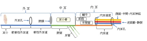
・外耳道とは 鼓膜 によって隔たれ、咽頭腔とは 耳管 で交通している。
 |
 |
 |
|
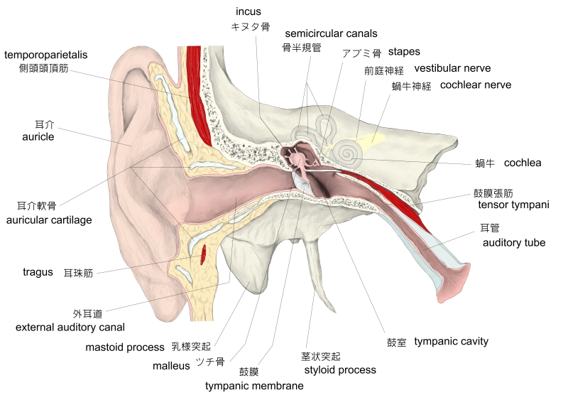
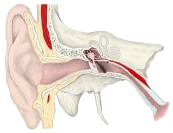
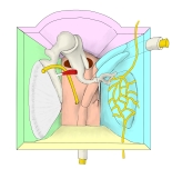
右耳・冠状断面・前面 |
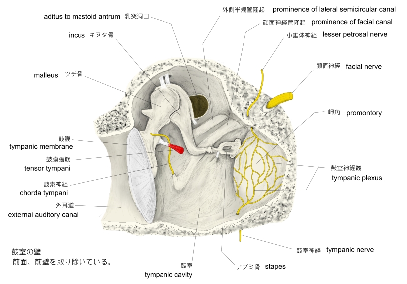
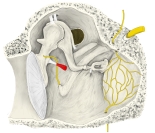
鼓室・前面 |
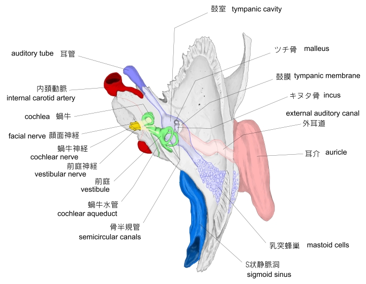
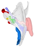
右側頭骨・前面 |
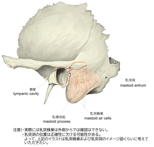
左側頭骨(外側面) |
「日本人体解剖学 (下巻) 」には以下のような解説が見られる。
■ 頭蓋底の中頭蓋窩の骨折 ■
「交通事故などで鼓室蓋の骨折があり、これに伴って鼓膜破裂や骨性外耳道の骨折が起こる。もし、脳硬膜が破れると多量の脳脊髄液が流出する。また、鼓室はしばしば疾病の温床といわれ、ここに起こった炎症はいろいろな部位に波及することが多い。」

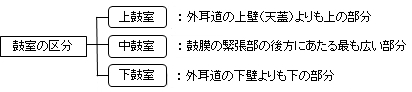
鼓室は上・中・下の3部に区分される。
「日本人体解剖学 (下巻) 」には以下のような解説が見られる。
「上鼓室:外耳道の上壁(天蓋)よりも上の狭い部分で、鼓膜の弛緩部の後上方にあたる。とくに、鼓膜の弛緩部に接するところをPrussak腔という。中耳炎と関係する重要な部位。 」
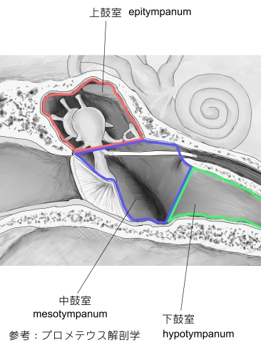
なお、「プロメテウス解剖学アトラス」では以下のように鼓室を3部に分けている。
・上鼓室(鼓室上陥凹): 鼓膜の高さより上方にある。
・中鼓室 : 鼓膜の内側にある。
・下鼓室(鼓室下陥凹): 鼓膜の高さより下方にある。
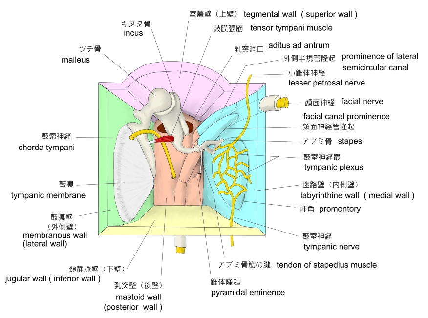
鼓室を取り囲む骨壁は、内側壁(迷路壁)、外側壁(鼓膜壁)、上壁(室蓋壁)、下壁、前、そして後の6つに区分することができる。以下は「日本人体解剖学 (下巻) 」を参考にしたものになる。
 |
|
|
|
右鼓室模型図(前面から見て) |
|
|
|

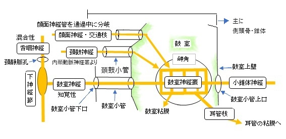
以下、鼓室に分布する神経、脈管を挙げてみたが、そのすべてを挙げているかどうかは不明となる。
【神 経】
1. 鼓室神経 : 下神経節から分岐、鼓室神経叢を形成、鼓室粘膜に分布、鼓室小管上口を出て小錐体神経となる。
2. 頚鼓神経 :内頚動脈神経叢より分岐、頚鼓小管を通って鼓室へ入り鼓室神経叢に合する。
3. 顔面神経交通枝 :顔面神経が顔面神経管を通過中に分岐、鼓室へ入り鼓室神経叢に合する。
※鼓索神経:この神経は鼓室は通過するのみと思われる。分布)舌神経に味覚線維、顎下腺・舌下腺に副交感性分泌線維
鼓索神経小管から鼓室に入り、鼓膜の内面でキヌタ骨とツチ骨の間を進み錐体鼓室裂から鼓室を出る。
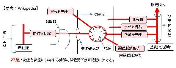
【動 脈】
1. 前鼓室動脈 : 顎動脈の第1区から分岐、鼓膜で分岐して血管輪を形成
2. 翼突管動脈:顎動脈の第4区から分岐、翼突管を後方に走る。
3. 内頚動脈の頚動脈鼓室枝
4. 茎乳突孔動脈が顔面神経管を通過中に分岐する以下の枝
a. 後鼓室動脈 b. アブミ骨枝 c. 乳突枝
【静 脈】
1.鼓室静脈:鼓索神経、前鼓室動脈とともに錐体鼓室裂を通過して鼓室から出て翼突筋静脈叢に注ぐ。
2.茎乳突孔静脈:鼓室から顔面神経管に入り茎乳突孔から出て翼突筋静脈叢へ。

The tympanic cavity is a small cavity surrounding the bones of the middle ear. Within it sit the ossicles, three small bones that transmit vibrations used in the detection of sound.
【Structure】
On its lateral surface, it abuts the external auditory meatus [ ear canal ] from which it is separated by the tympanic membrane (eardrum).
【Walls】
The tympanic cavity is bounded by:
- Facing the inner ear, the medial wall (or labyrinthic wall, labyrinthine wall) is vertical, and has the oval window and round window, the promontory, and the prominence of the facial canal.
- Facing the outer ear, the lateral wall (or membranous wall), is formed mainly by the tympanic membrane, partly by the ring of bone into which this membrane is inserted. This ring of bone is incomplete at its upper part, forming a notch (notch of Rivinus), close to which are three small apertures: the "iter chordæ posterius", the petrotympanic fissure, and the "iter chordæ anterius". The iter chordæ posterius (apertura tympanica canaliculi chordæ) is situated in the angle of junction between the mastoid and membranous wall of tympanic cavity immediately behind the tympanic membrane and on a level with the upper end of the manubrium of the malleus; it leads into a minute canal, which descends in front of the canal for the facial nerve, and ends in that canal near the stylo-mastoid foramen. Through it the chorda tympani nerve enters the tympanic cavity. The petrotympanic fissure opens just above and in front of the ring of bone into which the tympanic membrane is inserted; in this situation it is a mere slit about 2 mm. in length. It lodges the anterior process and anterior ligament of the malleus, and gives passage to the anterior tympanic branch of the internal maxillary artery. The iter chordæ anterius (canal of Huguier) is placed at the medial end of the petrotympanic fissure; through it the chorda tympani nerve leaves the tympanic cavity.
- The roof of the cavity (also called the tegmental wall, tegmental roof or tegmentum tympani) is formed by a thin plate of bone, the tegmen tympani, which separates the cranial and tympanic cavities. It is situated on the anterior (frontal) surface of the petrous portion of the temporal bone close to its angle of junction with the squama temporalis; it is prolonged backward so as to roof in the tympanic antrum, and forward to cover in the semicanal for the tensor tympani muscle. Its lateral edge corresponds with the remains of the petrosquamous suture.[1] The Atticus is the part of the tegmentum tympani where the stapes and incus are attached.
- The floor of the cavity (also called the jugular wall) is narrow, and consists of a thin plate of bone (fundus tympani) which separates the tympanic cavity from the jugular fossa. It presents, near the labyrinthic wall, a small aperture for the passage of the tympanic branch of the glossopharyngeal nerve.
- The posterior wall (or mastoid wall) is wider above than below, and presents for examination the entrance to the tympanic antrum, the pyramidal eminence, and the fossa incudis.
- The anterior wall (or carotid wall) is wider above than below; it corresponds with the carotid canal, from which it is separated by a thin plate of bone perforated by the tympanic branch of the internal carotid artery, and by the deep petrosal nerve which connects the sympathetic plexus on the internal carotid artery with the tympanic plexus on the promontory. At the upper part of the anterior wall are the orifice of the semicanal for the Tensor tympani muscle and the tympanic orifice of the auditory tube, separated from each other by a thin horizontal plate of bone, the septum canalis musculotubarius. These canals run from the tympanic cavity forward and downward to the retiring angle between the squama and the petrous portion of the temporal bone.
【Development】
It is formed from the tubotympanic recess, an expansion of the first pharyngeal pouch.
【 語 句 】
・ : ・ : ・ : ・ : ・ : ・ : ・ : ・ : ・ : ・ : ・ : ・ : ・ : ・ : ・ : ・ :

・イラストや写真を掲載しているサイト-Ⅰ
・イラストや写真を掲載しているサイト-Ⅱ
・イラストや写真を掲載しているサイト-Ⅲ
・イラストや写真を掲載しているサイト-Ⅳ(模型図)
・イラストや写真を掲載しているサイト-Ⅴ(模型図)