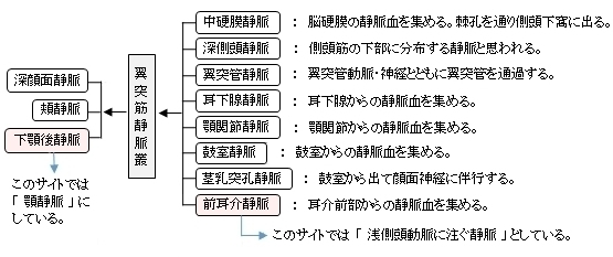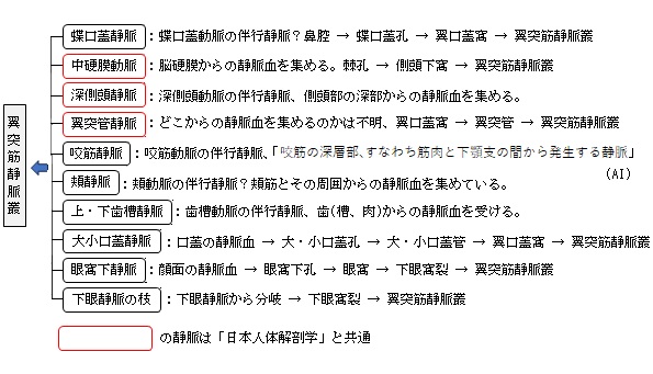


「 日本人体解剖学 」では以下のように、翼突筋静脈叢に注ぐ静脈として8つを、そして翼突筋静脈叢から出る静脈として3つを挙げている。(「 船戸和弥のHP 」でも、注ぐ静脈としては同じものを挙げていると思われる。)

以下は「Wikipedia」を参考にして作成したものとなる。


以下は「 Wikipedia 」の解説文となる。
「 The pterygoid plexus is a venous plexus of considerable size, and is situated between the temporalis muscle and lateral pterygoid muscle, and partly between the two pterygoid muscles.
【 Tributaries 】
It receives tributaries corresponding with the branches of the maxillary artery.
Thus it receives the following veins :
This plexus communicates freely with the anterior facial vein ; it also communicates with the cavernous sinus, by branches through the foramen Vesalii, foramen ovale, and foramen lacerum. Due to its communication with the cavernous sinus, infection of the superficial face may spread to the cavernous sinus, causing cavernous sinus thrombosis. Complications may include edema of the eyelids, conjunctivae of the eyes, and subsequent paralysis of cranial nerves which course through the cavernous sinus.
The pterygoid plexus of veins becomes the maxillary vein. The maxillary vein and the superficial temporal vein later join to become the retromandibular vein. The posterior branch of the retromandibular vein and posterior auricular vein then form the external jugular vein, which empties into the subclavian vein. 」
【 語 句 】
・anterior facial vein:=facial vein 顔面静脈 ・cavernous sinus:海綿静脈洞 ・foramen Vesalii:蝶形導出静脈孔 ・foramen ovale:卵円孔 ・foramen lacerum:破裂孔 ・cavernous sinus thrombosis:海綿静脈洞血栓症 ・complication:合併症 ・edema:浮腫・conjunctivae:結膜 ・ subsequent:続いて起こる ・paralysis:麻痺 ・superficial temporal vein:浅側頭静脈 ・retromandibular vein:下顎後静脈 ・posterior auricular vein:後耳介静脈 ・external jugular vein:外頚静脈 ・subclavian vein:鎖骨下静脈
【 イラスト掲載サイト 】
・ イラストや写真を掲載しているサイト-Ⅰ
・ イラストや写真を掲載しているサイト-Ⅱ
・ イラストや写真を掲載しているサイト-Ⅲ
・ イラストや写真を掲載しているサイト-Ⅳ
・ イラストや写真を掲載しているサイト-Ⅴ

|