内頚動脈とは

 |
 |
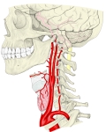 |
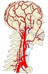
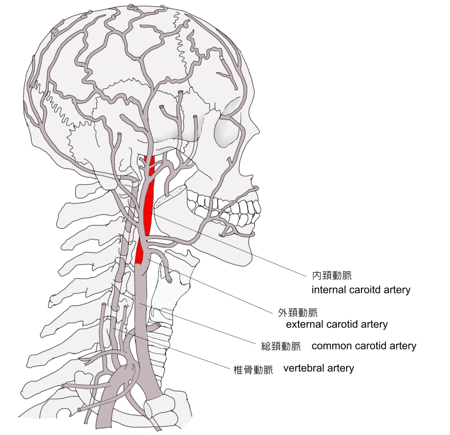
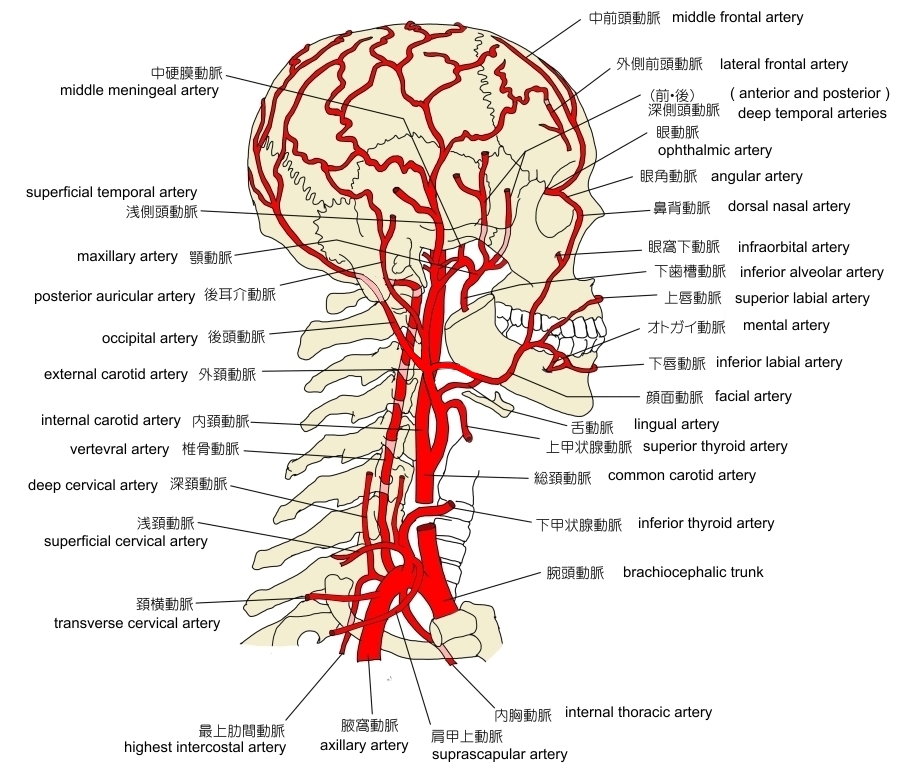
頚部・頭部の動脈
|
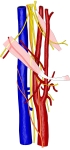
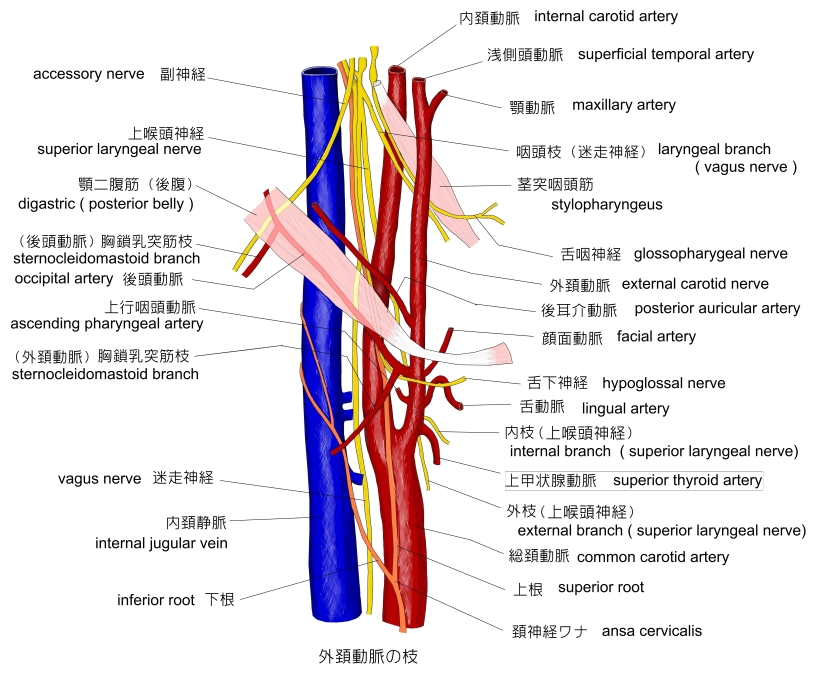
外頚動脈の枝 |
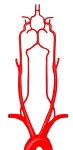
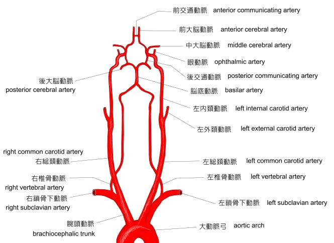
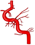
総頚動脈~前大脳動脈・模型図
|
|
|
|
|

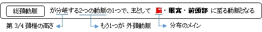
1 . 総頚動脈より分岐する。( 甲状軟骨の上縁/第4頚椎の高さで、外頚動脈の後内側に位置 )
2 . 軽いカーブは描くがほぼ垂直に咽頭の外側壁( 深頚筋の前側 )沿いを上行する。
(「外頚動脈と内頚動脈との間には茎突舌筋および茎突咽頭筋が経過する」( 日本人体解剖学))
3 . 頭蓋底に達する。
4 . 頚動脈管( 側頭骨岩様部 )に入り、次に頚動脈溝( 蝶形骨 )を海綿静脈洞に囲まれながら前進する。
5 . 視神経管の後方で眼動脈を分岐する。
6 . 脳硬膜を貫いて前/中大脳動脈となる。
以下は「船戸和弥のホームページ」の解説文となる。
「 局所解剖 : 頚部では内頚動脈は初めに外頚動脈の外側でやや後方にあるが,ついで外頚動脈の後方で内側にまがる.そこでは頭長筋と椎前筋膜のすぐそばにあり,また内側に接して咽頭がある.咽頭壁のそばを上行するときには,これと外頚動脈とのあいだに茎突舌筋と茎突咽頭筋がある.内頚静脈を伴っているが,この静脈は内頚動脈の後外側にあって頭蓋に達するのである.内頚動脈と内頚静脈の間でその後方を迷走神経が走り,さらに後内側に交感神経幹がある. 」

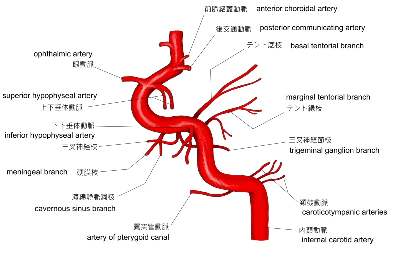
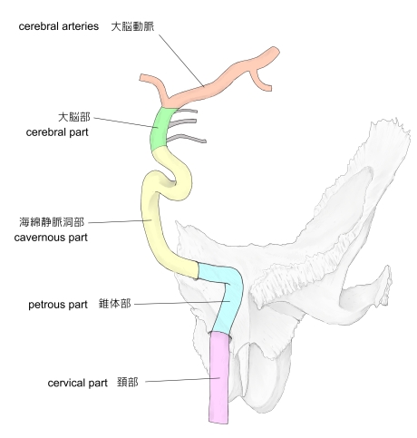
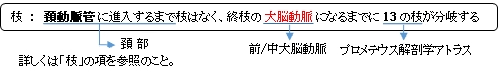
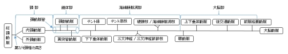
日本人体解剖学や船戸和弥のホームページのでは「区分」に関する言及は見られないが、プロメテウス解剖学アトラスでは、内頚動脈を以下の4部に区分している。以下の各部の解説文は全て「 プロメテウス解剖学アトラス:頭部/頚部解剖 」からの引用となる。
1 |
頚 部 |
cervical part 咽頭側隙に存在する。 |
2 |
錐体部 |
petrous part 側頭骨岩様部の頚動脈管に存在する。 |
3 |
海綿静脈洞部 |
cavernous part 海綿静脈洞のS状のカーブに存在する。 |
4 |
大脳部 |
cerebral part クモ膜下腔の交叉槽に存在する。 |
また「 プロメテウス解剖学アトラス:頭部/頚部解剖 」には以下のような解説文も見られる。
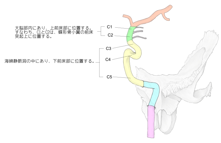
「 内頚動脈の頭蓋骨部は臨床的基準から5つの部分(C1-C5)に分けられる。」
・ C1-C2 : 大脳部内にあり、上前床部に位置する。すなわちC1とC2は、蝶形骨小翼の前床突起の上に位置する。
・ C3-C5 : 海綿静脈洞の中にあり、下前床部に位置する。

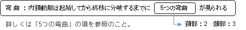
内頚動脈には、総頚動脈から起始してから終枝に分かれるまで、頚部に2つ、そして頭部に3つの弯曲が見られる。
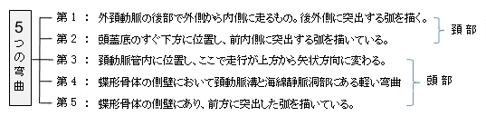
以下は「 日本人体解剖学 」からの引用文となる。
「 これらの2つの頚部湾曲を合わせると逆のS字形となる。」
「 これらの弯曲およびこの動脈の通る道がよく保護されていることは脳や眼に血流を供給する機構に重要な意味がある。」

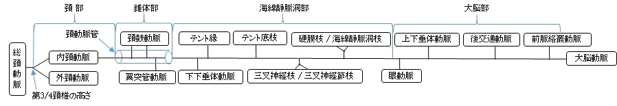
「 日本人体解剖学 」では、内頚動脈の枝に関しては眼動脈と大脳動脈の2つの動脈のみの解説が見られるだけとなる。それに対してプロメテウス解剖学アトラスでは、「 頭部で脳以外に分布する枝 」として区ごとに以下の枝を挙げている。
・「 内頚動脈は頚動脈管中で小枝、とくに頚動脈鼓室枝(頚鼓小管中を通る)を鼓室粘膜に与える。」( 日本人体解剖学 )
以下は内頚動脈の枝を区分ごとに簡単に表した図となる。※参考:プロメテウス解剖学アトラス

以下は「 Wikipedia 」の解説文となる。
「 The internal carotid artery is a major paired artery, one on each side of the head and neck, in human anatomy. They arise from the common carotid arteries where these bifurcate into the internal and external carotid arteries at cervical vertebral level 3 or 4; the internal carotid artery supplies the brain, while the external carotid nourishes other portions of the head, such as face, scalp, skull, and meninges.
Terminologia Anatomica in 1998 subdivided the artery into four parts: "cervical", "petrous", "cavernous", and "cerebral".[1][2]However, in clinical settings, the classification system of the internal carotid artery usually follows the 1996 recommendations by Bouthillier,[3] describing seven anatomical segments of the internal carotid artery, each with a corresponding alphanumeric identifier—C1 cervical, C2 petrous, C3 lacerum, C4 cavernous, C5 clinoid, C6 ophthalmic, and C7 communicating. The Bouthillier nomenclature remains in widespread use by neurosurgeons, neuroradiologists and neurologists. The segments are subdivided based on anatomical and microsurgical landmarks and surrounding anatomy, more than angiographic appearance of the artery. An alternative embryologic classification system proposed by Pierre Lasjaunias [4] and colleagues is invaluable when it comes to explanation of many internal carotid artery variants. An older clinical classification, based on pioneering work by Fischer,[5] is mainly of historical significance.
The segments of the internal carotid artery are as follows:
- ・ Cervical segment, or C1, identical to the commonly used Cervical portion
- ・ Petrous segment, or C2
- ・ Lacerum segment, or C3
- ・ Cavernous segment, or C4, almost identical to the commonly used Cavernous portion
- ・ Clinoid segment, or C5. This segment is not identified in some earlier classifications and lies between the commonly used cavernous portion and cerebral or supraclinoid portion.
- ・ Ophthalmic, or supraclinoid segment, or C6
- ・ Communicating, or terminal segment, or C7
The internal carotid artery is a terminal branch of the common carotid artery; it arises around the level of the fourth cervical vertebra when the common carotid bifurcates into this artery and its more superficial counterpart, the external carotid artery.
■ C1 : Cervical segment ■
The cervical segment, or C1, or cervical part of the internal carotid, extends from the carotid bifurcation until it enters the carotid canal in the skull anterior to the jugular foramen.
At its origin, the internal carotid artery is somewhat dilated. This part of the artery is known as the carotid sinus or the carotid bulb. The ascending portion of the cervical segment occurs distal to the bulb, when the vessel walls are again parallel.
The internal carotid runs vertically upward in the carotid sheath and enters the skull through the carotid canal. During this part of its course, it lies in front of the transverse processes of the upper three cervical vertebrae.
It is relatively superficial at its start, where it is contained in the carotid triangle of the neck, and lies behind and medial to the external carotid, overlapped by the sternocleidomastoid muscle, and covered by the deep fascia, the platysma, and integument: it then passes beneath the parotid gland, being crossed by the hypoglossal nerve, the digastric muscle and the stylohyoid muscle, the occipital artery and the posterior auricular artery. Higher up, it is separated from the external carotid by the styloglossus and stylopharyngeus muscles, the tip of the styloid process and the stylohyoid ligament, the glossopharyngeal nerve and the pharyngeal branch of the vagus nerve. It is in relation, behind, with the longus capitis, the superior cervical ganglion of the sympathetic trunk, and the superior laryngeal nerve; laterally, with the internal jugular vein and vagus nerve, the nerve lying on a plane posterior to the artery; medially, with the pharynx, superior laryngeal nerve, and ascending pharyngeal artery. At the base of the skull the glossopharyngeal, vagus, accessory, and hypoglossal nerves lie between the artery and the internal jugular vein.
Unlike the external carotid artery, the internal carotid normally has no branches in the neck.
【 語 句 】
・ bifurcation : 分岐(点) ・ carotid canal : 頚動脈管 ・ jugular foramen : 頚静脈孔 ・ dilated : 膨らんだ ・ carotid sinus : 頚動脈洞 ・ transverse process : 横突起 ・ sternocleidomastoid muscle : 胸鎖乳突筋 ・platysma : 広頚筋 ・ integument : 外皮 ・ parotid gland : 耳下腺 ・ hypoglossal nerve : 舌下神経 ・ digastric muscle : 顎二腹筋 ・ stylohyoid muscle : 茎突舌骨筋 ・ styloglossus muscle : 茎突舌骨 ・ stylopharyngeus muscle : 茎突咽頭筋 ・ glossopharyngeal nerve : 舌咽神経 ・ vagus nerve : 迷走神経 ・ longus capitis : 頭長筋 ・ superior cervical ganglion : 上頚神経節 ・ sympathetic trunk : 交感神経幹 ・ pharynx : 咽頭
■ C2 : Petrous segment ■
The petrous segment, or C2, of the internal carotid is that which is inside the petrous part of the temporal bone. This segment extends until the foramen lacerum. The petrous portion classically has three sections: an ascending, or vertical portion; the genu, or bend; and the horizontal portion.
When the internal carotid artery enters the canal in the petrous portion of the temporal bone, it first ascends a short distance and then curves anteriorly and medially. The artery lies at first in front of the cochlea and tympanic cavity; from the latter cavity it is separated by a thin, bony lamella, which is cribriform in the young subject, and often partly absorbed in old age. Farther forward it is separated from the trigeminal ganglion by a thin plate of bone, which forms the floor of the fossa for the ganglion and the roof of the horizontal portion of the canal. Frequently this bony plate is more or less deficient, and then the ganglion is separated from the artery by fibrous membrane. The artery is separated from the bony wall of the carotid canal by a prolongation of dura mater, and is surrounded by a number of small veins and by filaments of the carotid plexus, derived from the ascending branch of the superior cervical ganglion of the sympathetic trunk.
The named branches of the petrous segment of the internal carotid artery are:
■ C3 : Lacerum segment ■
The lacerum segment, or C3, is a short segment that begins above the foramen lacerum and ends at the petrolingual ligament, a reflection of periosteum between the lingula and petrous apex (or petrosal process) of the sphenoid bone. The lacerum portion is still considered 'extra-dural', as it is surrounded by periosteum and fibrocartilage along its course. The vidian artery arises from the lacerum segment, though it is often too small to be angiographically visible. It is erroneously stated in several anatomy text books that the internal carotid artery passes through the foramen lacerum. This at best has only ever been a partial truth in that it passes through the superior part of the foramen on its way to the cavernous sinus. As such it does not traverse the skull through it. The inferior part of the foramen is actually filled with fibrocartilage. The broad consensus is that the internal carotid artery should not be described as travelling through the foramen lacerum.[6]
【 語 句 】
・ petrolingual ligament : ? ・ reflection : 反映、投影? ・ periosteum : 骨膜 ・ lingula : 小舌 ・ petrous apex : 錐体尖? ・ fibrocartilage : 繊維軟骨 ・ Vidian artery : ヴィディウス動脈 ・ ngiographically : 管造影的に ・ cavernous sinus : 海綿静脈洞
■ C4 : Cavernous segment ■
The cavernous segment, or C4, of the internal carotid artery begins at the petrolingual ligament and extends to the proximal dural ring, which is formed by the medial and inferior periosteum of the anterior clinoid process. The cavernous segment is surrounded by the cavernous sinus.
In this part of its course, the artery is situated between the layers of the dura mater forming the cavernous sinus, but covered by the lining membrane of the sinus. It at first ascends toward the posterior clinoid process, then passes forward by the side of the body of the sphenoid bone, again curves upward on the medial side of the anterior clinoid process, and perforates the dura mater forming the roof of the sinus. The curve in the cavernous segment is called the carotid siphon. This portion of the artery is surrounded by filaments of the sympathetic trunk and on its lateral side is the abducent nerve, or cranial nerve VI.
The named branches of the cavernous segment are:
The cavernous segment also gives rise to small capsular arteries that supply the wall of the cavernous sinus.
【 語 句 】
・ petrolingual ligament: ・ dural ring :硬膜輪? ・ anterior clinoid process:前床突起 ・perforate :貫通する ・ carotid siphon:頚動脈サイホン ・ abducent nerve:外転神経
■ C5 : Clinoid segment ■
The clinoid segment, or C5, is another short segment of the internal carotid that begins after the artery exits the cavernous sinus at the proximal dural ring and extends distally to the distal dural ring, after which the carotid artery is considered "intra-dural" and has entered the subarachnoid space.
The clinoid segment normally has no named branches, though the ophthalmic artery may arise from the clinoid segment.
■ C6 : Ophthalmic segment ■
The ophthalmic segment, or C6, extends from the distal dural ring, which is continuous with the falx cerebri, to the origin of the posterior communicating artery. The ophthalmic segment courses roughly horizontally, parallel to the optic nerve, which runs superomedially to the carotid at this point.
The named branches of the ophthalmic segment are:
■ C7 : Communicating segment ■
The communicating segment, or terminal segment, or C7, of the internal carotid artery passes between the optic and oculomotor nerves to the anterior perforated substance at the medial extremity of the lateral cerebral fissure. Angiographically, this segment extends from the origin of the posterior communicating artery to the bifurcation of the internal carotid artery.
The named branches of the communicating segment are:
The internal carotid then divides to form the anterior cerebral artery and middle cerebral artery. The internal carotid artery can receive blood flow via an important collateral pathway supplying the brain, the cerebral arterial circle, which is more commonly known as the Circle of Willis.
【 語 句 】
・ oculomotor nerve : 動眼神経 ・ perforated : 貫通した ・ extremity : 末端 ・ lateral cerebral fissure : 外側大脳裂 ・collateral : 二次的な ・ cerebral arterial circle :大 脳動脈輪 ・ Circle of Willis : ウィリス動脈輪
【 branches 】
The following are the branches of the internal carotid artery, listed by segment:[7]
- ・ C1 : Branches from the cervical portion - none.
- ・ C2 : Branches from the petrous portion
- ・ C3 : Branches from the lacerum portion - none
- ・ C4 : Branches from the cavernous portion
- ・ C5 : Branches from the clinoid portion - none
- ・ C6 : Branches from the ophthalmic portion
- ・ C7 : Branches from the communicating portion
【 イラスト掲載サイト 】
・ イラストや写真を掲載しているサイト-Ⅰ
・ イラストや写真を掲載しているサイト-Ⅱ
・ イラストや写真を掲載しているサイト-Ⅲ
・ イラストや写真を掲載しているサイト-Ⅳ
・ イラストや写真を掲載しているサイト-Ⅴ

|