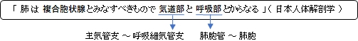
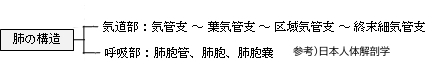
・概 要
・外見(肺尖、肺底、面、縁)
・肺門・肺根
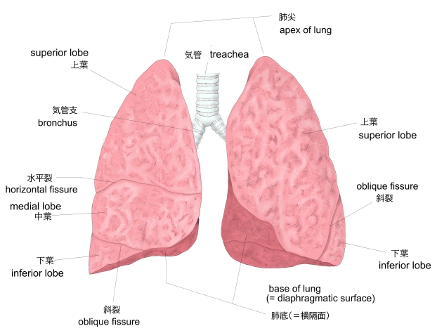
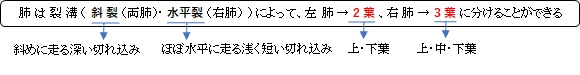
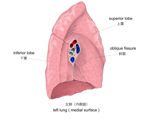
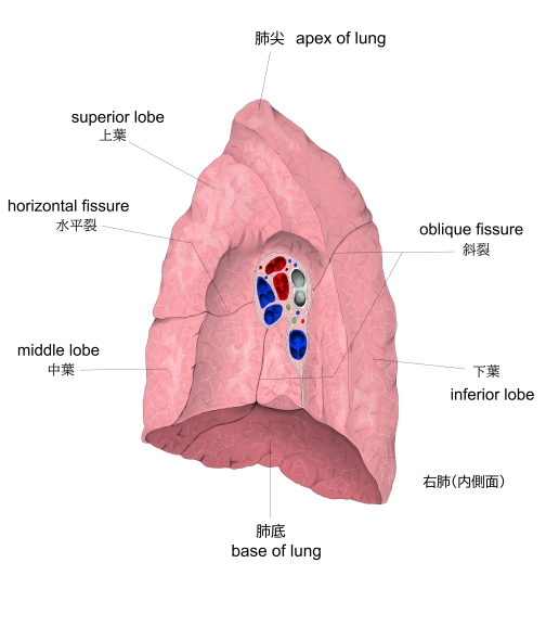
・肺 葉
・位 置
・気管支の分岐
・構 造
・脈管/神経
・発 生
・関連語句
・Wikipedia
・イラスト掲載サイト
![]()
 |
|
|||
胸 郭 |
肺(前面) |
「ウィキペディア」には以下のような解説文が見られる。
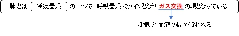
ヒトの場合、重さは両肺で男性が約1kg、女性は約0.9kgほどあり、胸腔の大部分を占め、
主に気道と血管からなり、ガス交換のため両者は肺胞で接している。肺胞は約3億個で、
総表面積は約70m2。

・「色調は真珠色が多い。」(日本人体解剖学)
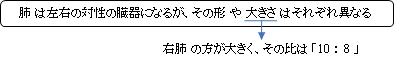
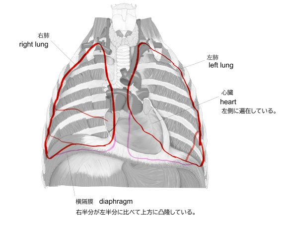
・「左肺は気管・食道・心臓および大動脈の左側、右肺はこれらの右側に位置する」(日本人体解剖学)
・「左肺は右肺よりも細長で、容積も小さく(左8:右10)、低位に位置する。」(日本人体解剖学)
・「重さは両肺で男性が約1kg、女性は約0.9kgほど」(ウィキペディア)
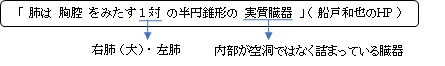
・「右肺(1200cc, 600g)は左肺(1100cc, 500g)よりやや大きい」(船戸和也のHP)
「日本人体解剖学」では、左右の肺の形状が異なる理由として以下を挙げている。
・心臓が左側に遍在している。
・肝臓の右葉が大きい。
・横隔膜の右半分が左半分に比べてより上方に凸隆している。
 |
||||
左肺・右肺 |
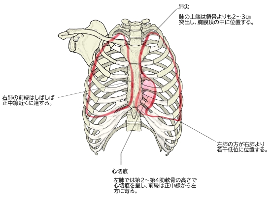
肺は上端と下底、そして面および線を区別する。
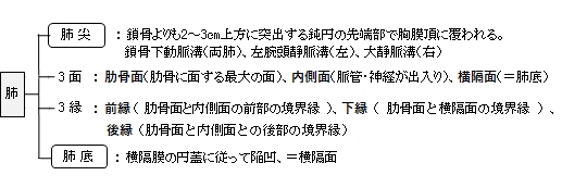
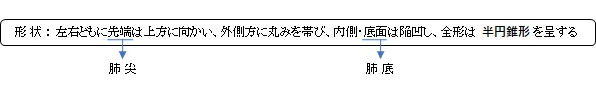
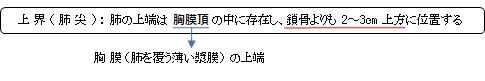
【 肺 尖 】
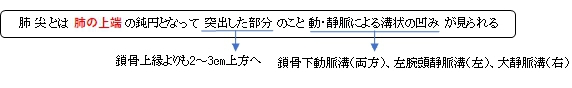
・「胸膜頂におおわれる」(日本人体解剖学)
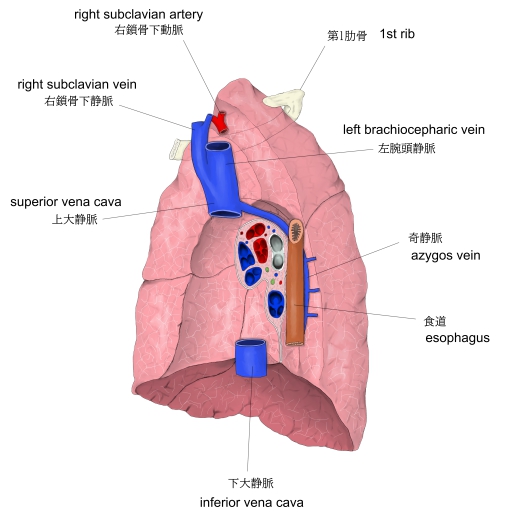
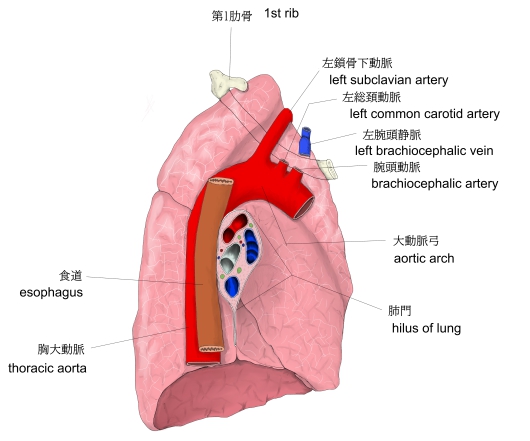
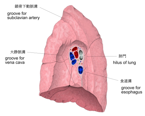
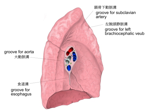
以下は「日本人体解剖学」の解説文となる。左右の肺尖の内側には鎖骨下動脈による鎖骨下動脈溝
がみられる。さらに、左肺は左腕頭静脈による左腕頭静脈溝、右肺は上大静脈による
大静脈溝を認める。
 |
 |
 |
 |
右肺(内側面) |
左肺(内側面) |
右肺の溝 |
左肺の溝
|
【 肺 底 】
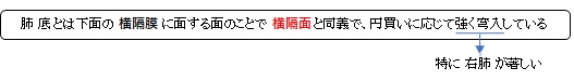
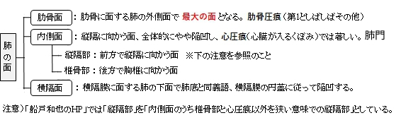
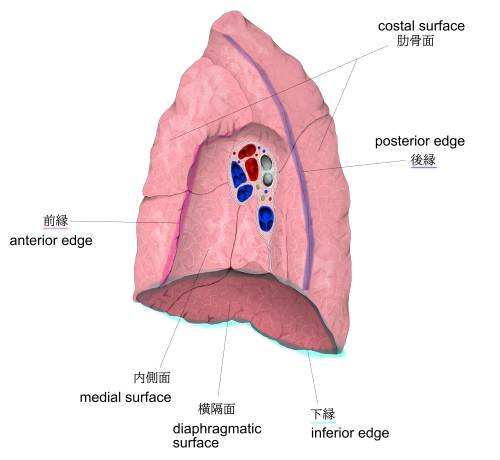
【 面 】
肺は以下の3つの面を区別する。
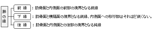
【 縁 】
肺は以下の3つの縁を区別する。3つとも面同士の境界縁となる。
|
 |
 |
||
左肺・右肺 |
肺の面 |
縁 |
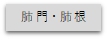
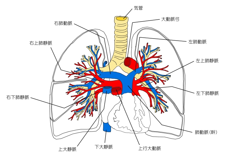
・「肺門では気管支が最も後方に、その前方に肺動脈、さらに前方に肺静脈が位置する。」(日本人体解剖学)
・「(肺門は)内側面の中央で、およそ第6胸椎の高さにある。」(船戸和也のHP)
・「肺門周辺にはリンパ節(気管支肺リンパ節)が発達し、肺内からのリンパを集める。」(船戸和也のHP)
![]()
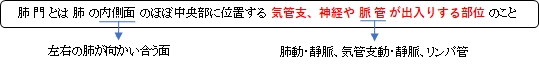
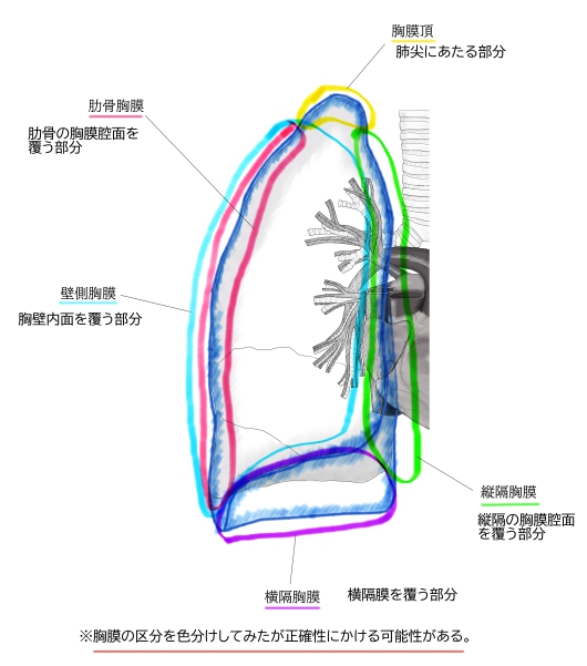
「船戸和也のHP」には以下のような解説文が見られる。
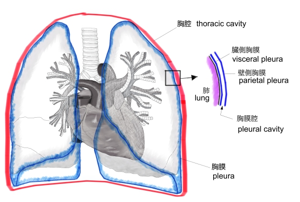
「肺門は、肺の表面をおおう肺胸膜(臓側胸膜)が肺根を包み胸腔壁をおおう壁側胸膜に移行するところで、
肺根は上・前・後の3面から肺胸膜によって覆われる。下方では前後の胸膜は肺門から遠ざかるにつれて次第
に接着し、肺門下部から横隔面に及ぶ二重葉となり、肺間膜(pulmonary ligament)と呼ばれる。」
 |
 |
|||
右肺の肺門 |
左肺の肺門 |

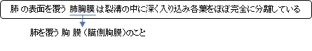
「日本人体解剖学」には以下のような解説文が見られる。
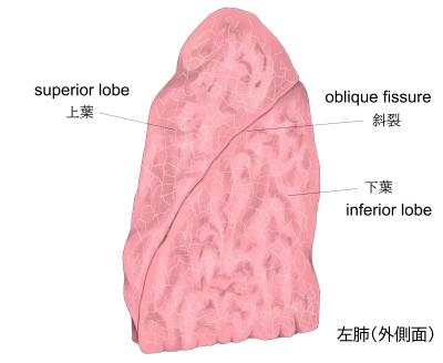
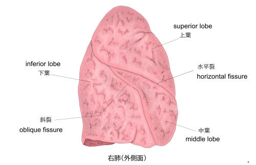
「肺の表面を覆う肺胸膜は、斜裂と水平裂の中に入り込み各葉をほとんど完全に分離し、
各葉はただ気管支枝によってのみ連絡される。」
|
 |
 |
 |
 |
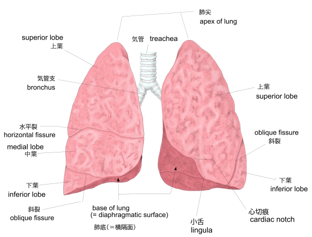
肺(前面) |
左肺(内側面) |
右肺(内側面) |
左肺(外側面) |
右肺(外側面) |

![]()
・「左肺は気管・食道・心臓および大動脈の左側、右肺はこれらの右側に位置する」(日本人体解剖学)
【肺の前界】
「日本人体解剖学」には以下のような解説文が見られる。
肺の前縁に一致して左右ともに胸鎖関節の後側から弧状を呈し他側のものに近づきつつ下方
に走り、第6肋軟骨の高さで肺の下界に移行する。ただし、左側では第2~第4肋軟骨の高さで
著明な心切痕を示して正中線から左方に遍在するが、右肺のものはしばしば正中線に達する。
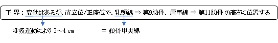
・「左肺は右肺よりも細長で、容積も小さく(左8:右10)、低位に位置する。」(日本人体解剖学)
 |
 |
 |
 |
|
胸郭と肺(前面) |
体幹に垂直な 参照線(前面) |
体幹に垂直な 参照線(後面) |
胸膜の区分 |
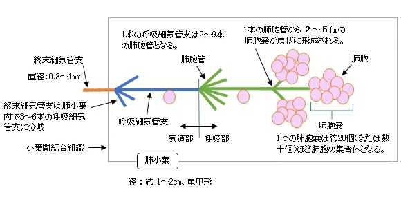
以下は「日本人体解剖学」の解説文となる。
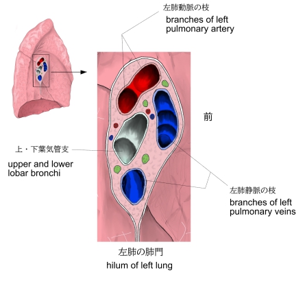
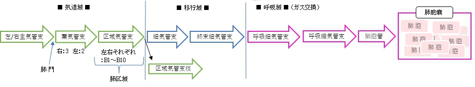
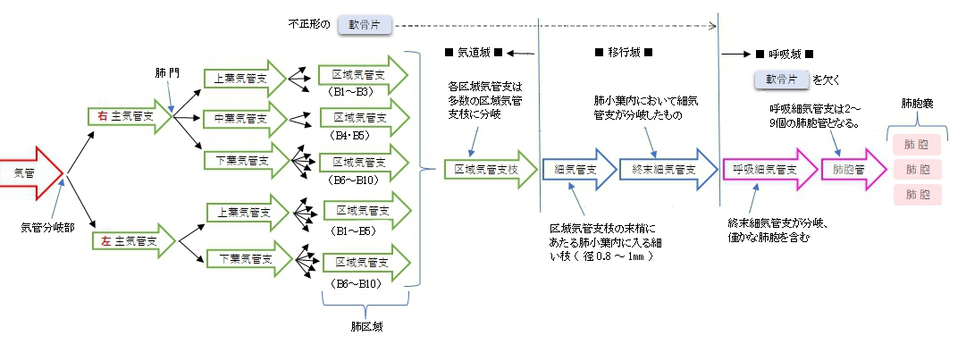
「 左、右の気管支は次のように分岐していく。
葉気管支→区域気管支→細気管支→終末細気管支→呼吸細気管支→肺胞管→ 肺胞嚢→肺胞 」
1 |
lober bronchus |
肺門において(主)気管支から分岐する(左2本、右3本)。肺実質内に入り、構造的には気管支をほぼ同一となるが、異なる点もいくつか見られる。 特徴) ・膜性壁を欠く。 ・C字形の軟骨片は次第に退縮 ⇒ 不正形の軟骨片(径1㎜大の枝にまで見られる) ・多列線毛円柱上皮は次第に上皮細胞の丈が低くなる。 |
2 |
segmental bronchus |
|
3 |
bronchiole |
|
4 |
terminal bronchiole |
|
5 |
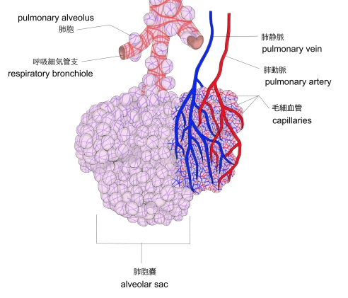
respiratory bronchiole |
![]()

気管に分布する脈管・神経として以下のものが挙げられる。
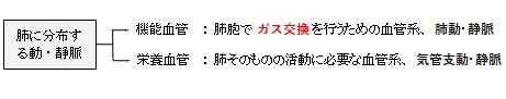
【機能血管】
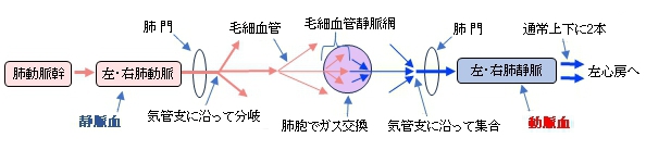
以下は「日本人体解剖学」の解説文となる。
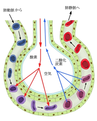
「肺動・静脈、静脈血を肺に運ぶ肺動脈は、左右1本ずつあって肺門から入り、気管支の
分岐に沿って分岐し肺胞を毛細血管網で囲む。ここでガス交換がされ、酸素濃度の高い
血液は静脈に入り、気管支に沿って走行しながら次第に集合して幹となり、ついに肺静脈
として肺門を出る。」
 |
 |
 |
|
肺動・静脈 |
肺胞嚢と動・静脈 |
ガス交換 |
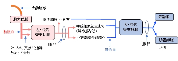
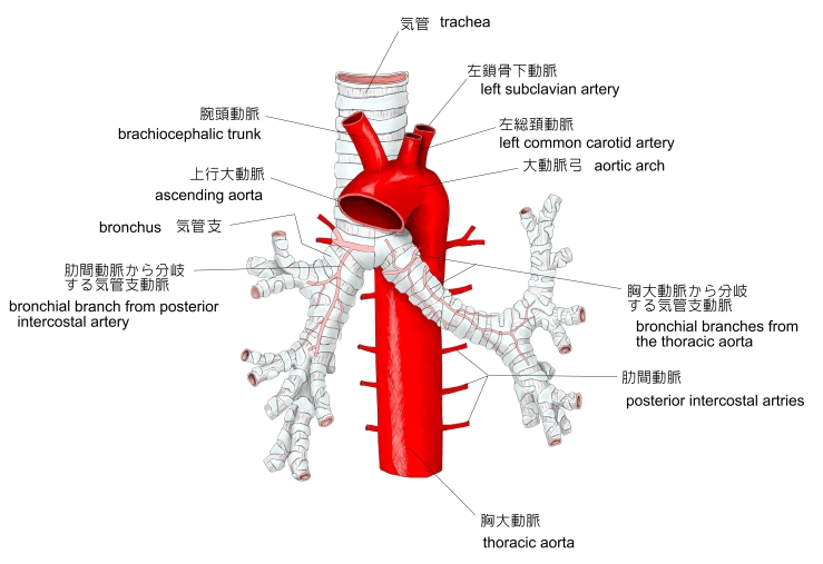
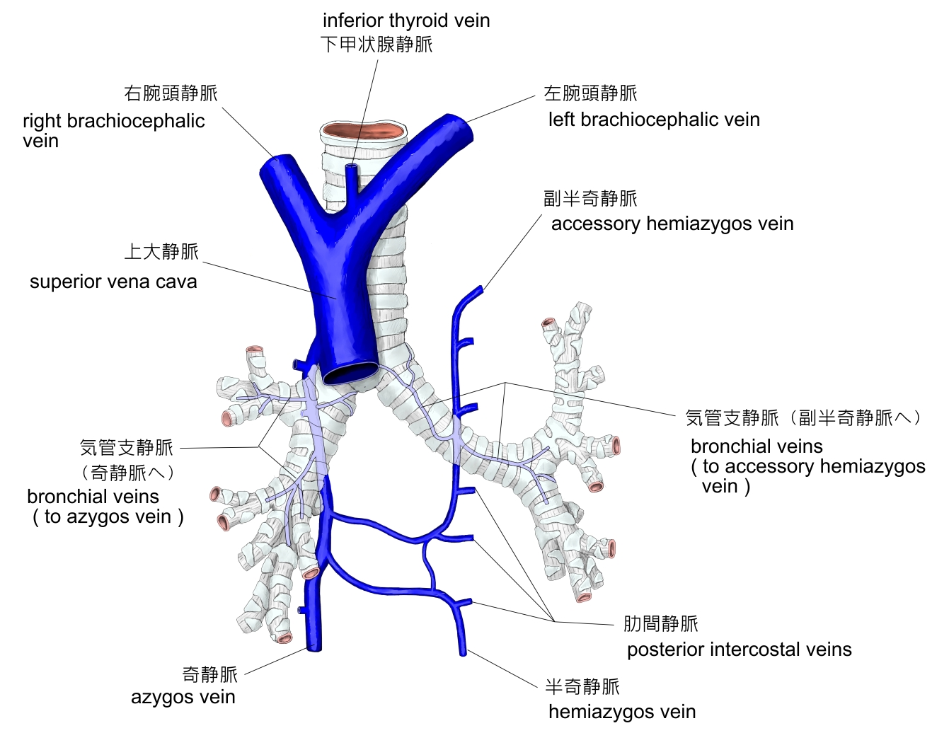
【栄養血管】
以下は「日本人体解剖学」の解説文となる。
「気管支動・静脈、気管支動脈は気管支分岐にほぼ随伴して走る。胸大動脈から2~3本あるいは
共通幹をもって起こり、左右に分かれ気管支に分布して筋・腺・肺の血管壁を養うほか胸膜にも分布
する。これから出た静脈血は気管支静脈となり、肺門を出て奇静脈または肋間静脈(左側)に入る。」
 |
 |
||||
気管支動脈 |
気管支静脈 |
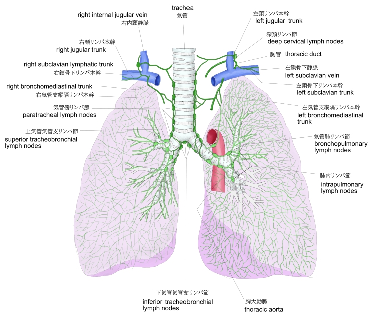
【リンパ管】
以下は「日本人体解剖学」の解説文となる。
肺表面のものは肺胸膜下リンパ叢となり、気管支リンパ節、肺リンパ節に入る。末梢部のものは肺胞、肺胞管および気管支のまわりに分布し、肺門にある気管支肺リンパ節に入る。
 |
||||
気管支・肺 |
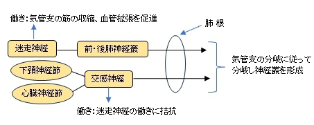
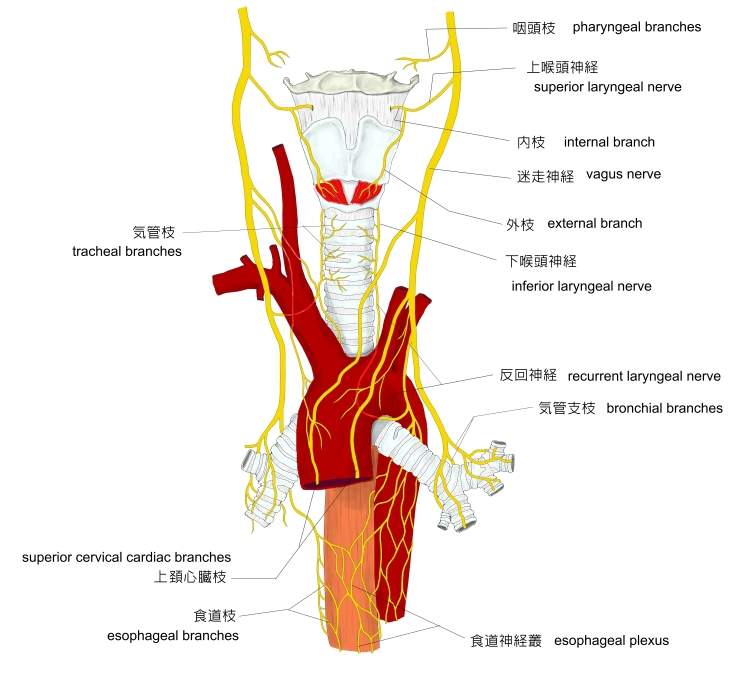
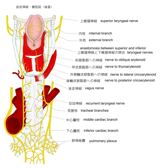
【神 経】
以下は「日本人体解剖学」の解説文となる。
迷走神経および交感神経が分布し、肺門から気管支の分岐に従って分岐し神経叢をつくる。迷走神経は気管支の筋の収縮と血管拡張を促し、交感神経はこれに拮抗する。
迷走神経は前・後肺神経叢として肺根に入り、下頚神経節および心臓神経節からの交感神経線維もまたここに入る。なお、気管支の分岐に沿って交感神経細胞群がみられることがある。
 |
 |
 |
||||
神経-1 |
神経-2 |
自律神経 |
![]()
以下は「日本人体解剖学」の「気管および肺の発生」の解説文となる。
前腸の前壁にできた呼吸器原基をなす盲嚢の末端は、胎生第4週の終わりに分岐して気管支をつくるが、この際、右側は3分し、左側は2分して(完成)後の肺葉数を示す。ついで第二次分岐を繰り返し、その末端部は膨大して細気管支、終末細気管支および原始肺胞はまた肺胞管となる。胎生末期になると肺胞の発生が特に著しくなり、呼吸細気管支から肺胞が膨出する。そして、生後には肺胞は新生は行われない。
肺胞の上皮は胎生末期まで線毛をもつが、その後は線毛を失い、生後空気を受容するようになると肺胞管は拡大して、肺胞壁の上皮も扁平上皮と変わる。肺の中の結合組織、弾性線維および軟骨は中胚葉からできる。

か行 |
は行 | ||
| 奇静脈葉 | |||
| さ行 | |||
| 左腕頭静脈溝 | |||
| 副葉 | |||
| た行 | や行 | ||

以下は「Wikipedia」の解説文となる。
The lungs are the central organs of the respiratory system in humans and some other animals, including tetrapods, some snails and a small number of fish. In mammals and most other vertebrates, two lungs are located near the backbone on either side of the heart. Their function in the respiratory system is to extract oxygen from the air and transfer it into the bloodstream, and to release carbon dioxide from the bloodstream into the atmosphere, in a process of gas exchange. The pleurae, which are thin, smooth, and moist, serve to reduce friction between the lungs and chest wall during breathing, allowing for easy and effortless movements of the lungs.
Respiration is driven by different muscular systems in different species. Mammals, reptiles and birds use their different muscles to support and foster breathing. In earlier tetrapods, air was driven into the lungs by the pharyngeal muscles via buccal pumping, a mechanism still seen in amphibians. In humans, the main muscle of respiration that drives breathing is the diaphragm. The lungs also provide airflow that makes vocal sounds including human speech possible.
Humans have two lungs, one on the left and one on the right. They are situated within the thoracic cavity of the chest. The right lung is bigger and heavier than the left, which shares space in the chest with the heart. The lungs together weigh approximately 1.3 kilograms (2.9 pounds). The lungs are part of the lower respiratory tract that begins at the trachea and branches into the bronchi and bronchioles, and which receive air breathed in via the conducting zone. The conducting zone ends at the terminal bronchioles. These divide into the respiratory bronchioles of the respiratory zone which divide into alveolar ducts that give rise to the alveolar sacs that contain the alveoli, where gas exchange takes place. Alveoli are also sparsely present on the walls of the respiratory bronchioles and alveolar ducts. Together, the lungs contain approximately 2,400 kilometres (1,500 miles) of airways and 300 to 500 million alveoli. Each lung is enclosed within a pleural sac of two membranes called pleurae; the membranes are separated by a film of pleural fluid, which allows the inner and outer membranes to slide over each other whilst breathing takes place, without much friction. The inner pleura also divides each lung into sections called lobes. The right lung has three lobes and the left has two. The lobes are further divided into bronchopulmonary segments and pulmonary lobules. The lungs have a unique blood supply, receiving deoxygenated blood from the heart in the pulmonary circulation for the purposes of receiving oxygen and releasing carbon dioxide, and a separate supply of oxygenated blood to the tissue of the lungs, in the bronchial circulation. Deoxygenated blood travels from the heart through the pulmonary artery to the lungs to be oxygenated in capillaries of alveoli. After the blood is oxygenated, it returns to the heart through the pulmonary vein to be sent to the rest of the body.[1][2]
The tissue of the lungs can be affected by a number of respiratory diseases, including pneumonia and lung cancer. Chronic obstructive pulmonary disease includes chronic bronchitis and emphysema, and can be related to smoking or exposure to harmful substances. A number of occupational lung diseases can be caused by substances such as coal dust, asbestos fibres, and crystalline silica dust. Diseases such as bronchitis can also affect the respiratory tract. Medical terms related to the lung often begin with pulmo-, from the Latin pulmonarius (of the lungs) as in pulmonology, or with pneumo- (from Greek πνεύμων "lung") as in pneumonia.
In embryonic development, the lungs begin to develop as an outpouching of the foregut, a tube which goes on to form the upper part of the digestive system. When the lungs are formed the fetus is held in the fluid-filled amniotic sac and so they do not function to breathe. Blood is also diverted from the lungs through the ductus arteriosus. At birth, however, air begins to pass through the lungs, and the diversionary duct closes, so that the lungs can begin to respire. The lungs only fully develop in early childhood.
【語句】
・: ・: ・: ・: ・: ・: ・: ・: ・: ・: ・: ・: ・: ・: ・: ・: ・: ・: ・: ・: ・: ・: ・: ・: ・: ・: ・: ・: ・: ・: ・: ・: ・: ・: ・: ・:
■ 写真やイラストを掲載しているサイト ■
![]()