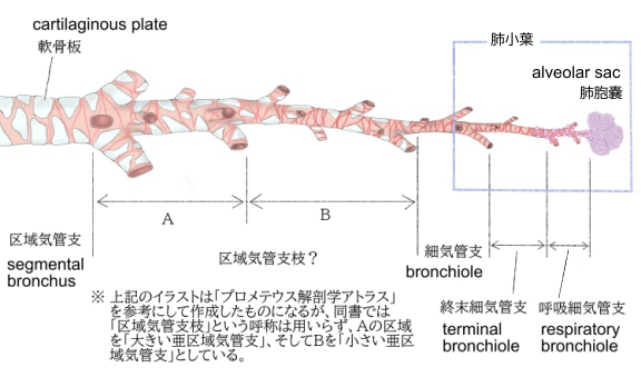
1. 直径は0.8~1㎜
2. 1本の細気管支の分布する直径1~2㎝の円錐形の範囲を肺小葉 という。
3. 肺小葉内で3~6本に分岐し次の枝に移行
4. 太い部分の細気管支の上皮は胚細胞(germ cell)をまじえた多列繊毛上皮(pseudostratified ciliated epithelium)
5. 固有層には気管支腺(混合腺)が存在
6. 固有層の下には平滑筋が走る。
7. 筋層の下には疎性結合組織の層があり軟骨片が見られる。
8. 直径が1㎜以下になると軟骨は見られず平滑筋が発達して輪走する。
以下は「Wikipedia」の解説文となる。
The bronchioles or bronchioli (pronounced bron-kee-oh-lee) are the smaller branches of the bronchial airways in the lower respiratory tract. They include the terminal bronchioles, and finally the respiratory bronchioles that mark the start of the respiratory zone delivering air to the gas exchanging units of the alveoli. The bronchioles no longer contain the cartilage that is found in the bronchi, or glands in their submucosa.[1]
Structure
The pulmonary lobule is the portion of the lung ventilated by one bronchiole. Bronchioles are approximately 1 mm or less in diameter and their walls consist of ciliated cuboidal epithelium and a layer of smooth muscle. Bronchioles divide into even smaller bronchioles, called terminal, which are 0.5 mm or less in diameter. Terminal bronchioles in turn divide into smaller respiratory bronchioles which divide into alveolar ducts. Terminal bronchioles mark the end of the conducting division of air flow in the respiratory system while respiratory bronchioles are the beginning of the respiratory division where gas exchange takes place.
The diameter of the bronchioles plays an important role in air flow. The bronchioles change diameter to either increase or reduce air flow. An increase in diameter is called bronchodilation and is stimulated by either epinephrine or sympathetic nerves to increase air flow. A decrease in diameter is called bronchoconstriction, which is the tightening of the smooth muscle surrounding the bronchi and bronchioles due to and stimulated by histamine, parasympathetic nerves, cold air, chemical irritants, excess mucus production, viral infections, and other factors to decrease air flow. Bronchoconstriction can result in clinical symptoms such as wheezing, chest tightness, and dyspnea, which are common features of asthma, chronic obstructive pulmonary disease (COPD), and chronic bronchitis. [2]
Bronchioles
The trachea divides into the left main bronchus which supplies the left lung, and the right main bronchus which supplies the right lung. As they enter the lungs these primary bronchi branch into secondary bronchi known as lobar bronchi which supply each lobe of the lung. These in turn give rise to tertiary bronchi (tertiary meaning "third"), known as segmental bronchi which supply each bronchopulmonary segment.[1] The segmentary bronchi subdivide into fourth order, fifth order and sixth order segmental bronchi before dividing into the bronchioles. The bronchioles are histologically distinct from the bronchi in that their walls do not have hyaline cartilage and they have club cells in their epithelial lining. The epithelium of the bronchioles starts as a simple ciliated columnar epithelium and changes to simple ciliated cuboidal epithelium as the bronchioles decreases in size. The diameter of the bronchioles is often said to be less than 1 mm, though this value can range from 5 mm to 0.3 mm. As stated, these bronchioles do not have hyaline cartilage to maintain their patency. Instead, they rely on elastic fibers attached to the surrounding lung tissue for support. The inner lining (lamina propria) of these bronchioles is thin with no glands present, and is surrounded by a layer of smooth muscle. As the bronchioles get smaller they divide into terminal bronchioles. Each bronchiole divides into between 50 and 80 terminal bronchioles.[3] These bronchioles mark the end of the conducting zone, which covers the first division through the sixteenth division of the respiratory tract. Alveoli only become present when the conducting zone changes to the respiratory zone, from the sixteenth through the twenty-third division of the tract.
【語句】
・: ・: ・: ・: ・: ・: ・: ・: ・: ・: ・: ・: ・: ・: ・: ・: ・: ・: ・: ・: ・: ・: ・: ・: ・: ・: ・: ・: ・: ・: ・: ・: ・: ・: ・: ・:
■ 写真やイラストを掲載しているサイト ■
・ イラストや写真を掲載しているサイト-Ⅰ
・ イラストや写真を掲載しているサイト-Ⅱ
・ イラストや写真を掲載しているサイト-Ⅲ