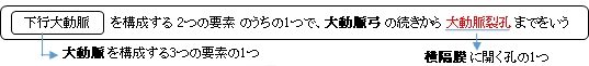
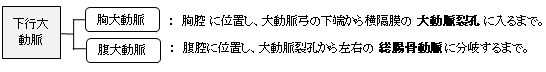
胸大動脈 とは


・「 半径は1.16㎝ほど 」( Wikipedia )


大動脈 |
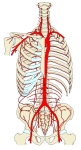
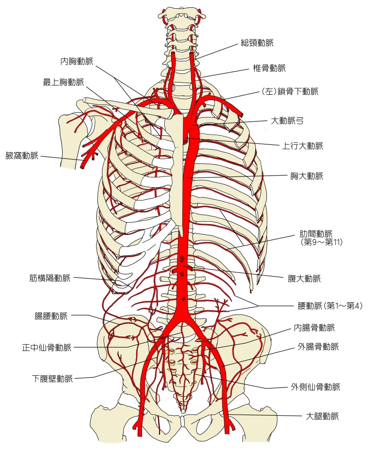
上半身の
主な動脈 |
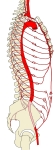
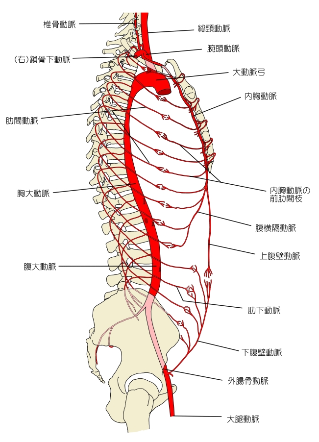
胸部腹部の
主な動脈 |
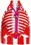
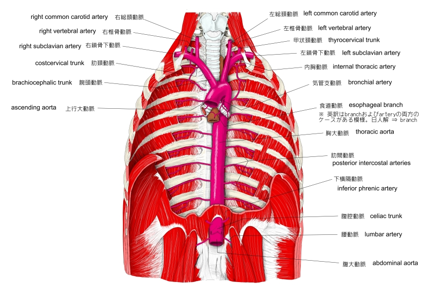
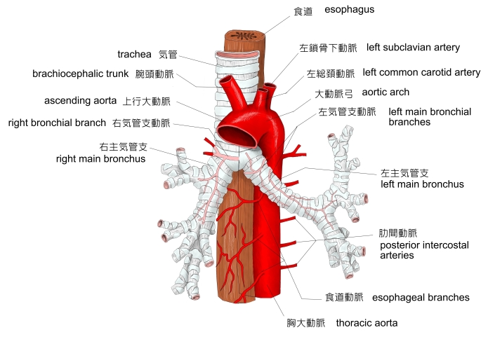
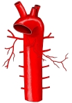
胸腔( 前面 )
⇒ 動脈以外の名称 |
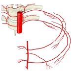
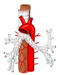
胸大動脈+
肋間動脈 |
大動脈弓~
胸大動脈 |
胸大動脈+
気管支+食道
|
|

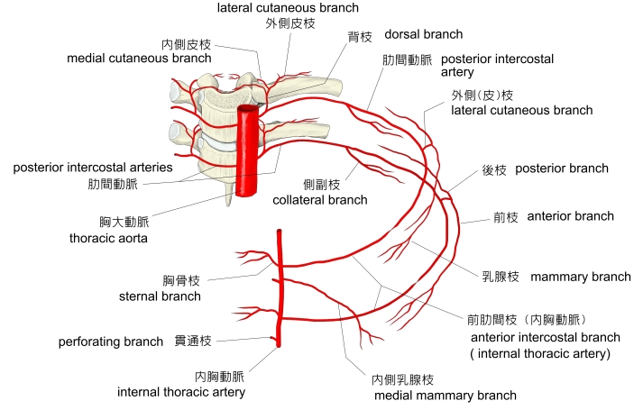
「 日本人体解剖学 」では、胸大動脈から分岐する枝を大きく臓側枝と壁側枝に区別しているが、「 船戸和弥のホームページ 」では、臓側枝および壁側枝という呼称は見られない。
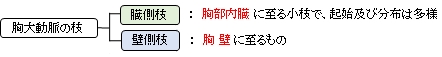
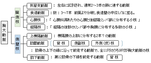
以下は胸大動脈の枝を簡単に表したものとなる。参考:「 日本人体解剖学 」

以下は「 Wikipedia 」の解説文となる。
「 The descending thoracic aorta is a part of the aorta located in the thorax. It is a continuation of the descending aorta and contained in the posterior mediastinal cavity. The descending thoracic aorta begins at the lower border of the fourth thoracic vertebra where it is continuous with the aortic arch, and ends in front of the lower border of the twelfth thoracic vertebra, at the aortic hiatus in the diaphragm where it becomes the abdominal aorta.
At its commencement, it is situated on the left of the vertebral column ; it approaches the median line as it descends; and, at its termination, lies directly in front of the column.
The descending thoracic aorta has a curved shape that faces forward, and has small branches. It has a radius of approximately 1.16 cm.[1]
【 語 句 】
・ thorax : 胸郭 ・ posterior mediastinal : 後縦隔 ・ aortic hiatus : 大動脈裂孔 ・ diaphragm : 横隔膜 ・ commencement : 開始 ・ vertebral column : 脊柱 ・ median line :正 中線 ・ radius : 半径
【 Structure 】
Main article: Aorta
The descending thoracic aorta is part of the aorta, which has different parts named according to their structure or location. The descending thoracic aorta is a continuation of the descending aorta and becomes the abdominal aorta when it passes through the diaphragm. The initial part of the aorta, the ascending aorta, rises out of the left ventricle, from which it is separated by the aortic valve. The two coronary arteries of the heart arise from the aortic root, just above the cusps of the aortic valve. The aorta then arches back over the right pulmonary artery. Three vessels come out of the aortic arch: the brachiocephalic artery, the left common carotid artery, and the left subclavian artery. These vessels supply blood to the head, neck, thorax and upper limbs.
Behind the descending thoracic aorta is the vertebral column and the hemiazygos vein. To the right is the azygos veins and thoracic duct, and to the left is the left pleura and lung. In front of the descending thoracic aorta lies the root of the left lung, the pericardium, the esophagus, and the diaphragm.
The esophagus, which is covered by a nerve plexus lies to the right of the descending thoracic aorta. Lower, the esophagus passes in front of the aorta, and ultimately is situated on the left.
【 語 句 】
・ left ventricle : 左心室 ・ aortic valve : 大動脈弁 ・ pulmonary artery : 肺動脈 ・ brachiocephalic artery : 腕頭動脈 ・ common carotid artery : 総頚動脈 ・ thorax : 胸部 ・ hemiazygos vein : 半奇静脈 ・ azygos veins : 奇静脈 ・ thoracic duct : 胸管 ・ pleura : 胸膜 ・ pericardium : 心膜 ・ esophagus : 食道 ・ ultimately : 最後には
【 Function 】
The aorta is an artery that conveys oxygenated blood from the heart to other parts of the body. It is one of the largest arteries in the body.[citation needed] The aorta gives off several paired branches as it descends. In descending order, these include the
Note : The posterior intercostal arteries are branches that originate throughout the length of the posterior aspect of the descending thoracic aorta. 」
【 語 句 】
・ posterior intercostal arteries : 肋間動脈
【 イラスト掲載サイト 】
・ イラストや写真を掲載しているサイト-Ⅰ
・ イラストや写真を掲載しているサイト-Ⅱ
・ イラストや写真を掲載しているサイト-Ⅲ
・ イラストや写真を掲載しているサイト-Ⅳ
・ イラストや写真を掲載しているサイト-Ⅴ

|