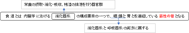
・概 要
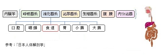
・区 分
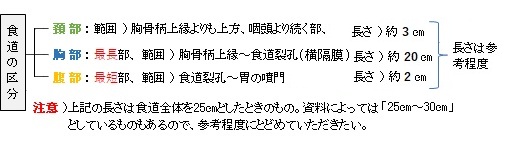
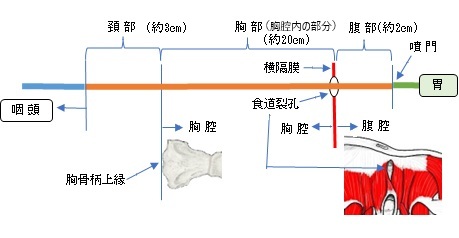
頚部、胸部、腹部
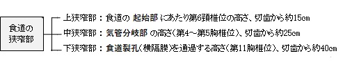
・生理的狭窄部
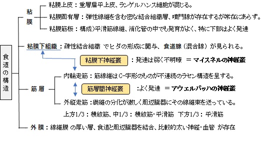
・構 造
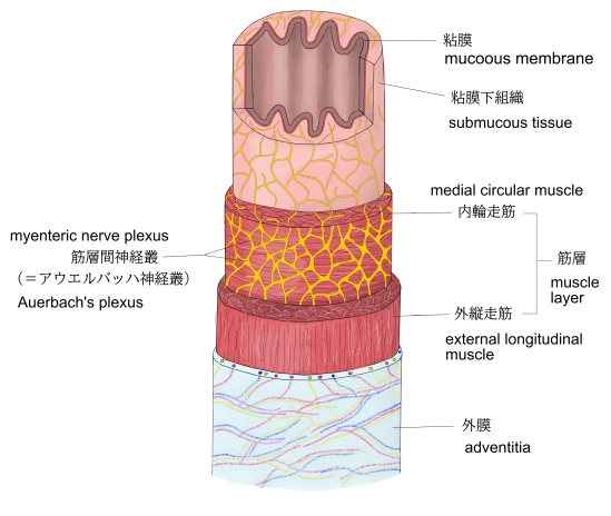
粘膜、粘膜下組織、筋層、外膜
・腺
・脈 管 / 神 経
・発 生
・Wikipedia
・イラストを掲載しているサイト
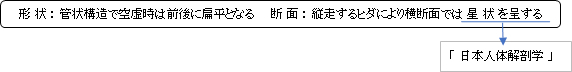
・「前後に扁平」という表現は「日本人体解剖学」を参考にしたものだが、インターネットで画像検索をかけると、イラストのほとんどは「左右に扁平」になっている。
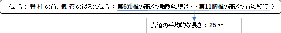
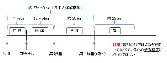
参 考 : 切歯~噴門( 37 ~ 40 cm)、切歯~食道(15cm) 「日本人体解剖学」 ・「長さ25cmぐらい、太さ2~3cm、厚さ約4mmのくだ状の臓器」(日本食道学会)
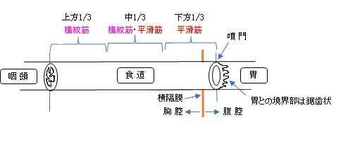
食道はその高さに従って3部構成(頚部、胸部、腹部)となる。 食道のそれぞれの長さをChatGPTに尋ねると以下のような回答であった。 頚部 (約10㎝~12㎝)、 胸部 (約15㎝~18㎝)、 腹部 (約2㎝~4㎝)
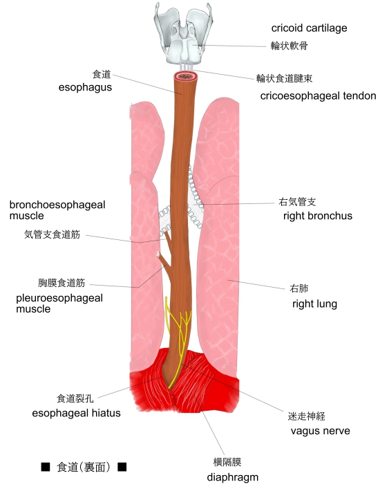
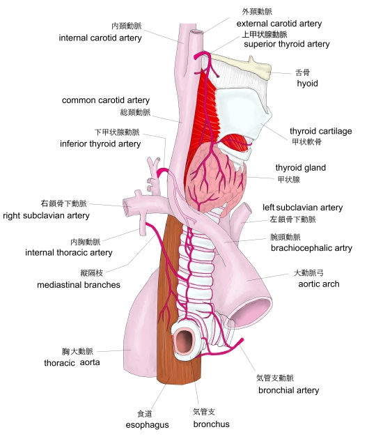
■ 頚 部:cervical part ■ ・胸骨柄上縁の高さにより上方に位置する短い部分。 ・気管、脊柱にはさまれてまっすぐ下行するが、下部ではやや左よりとなる。 ・気管とは疎性結合組織で結合し、その中あるいはやや外側を左・右反回神経が通る。 ・頚部の外側を左・右総頚動脈が通る。
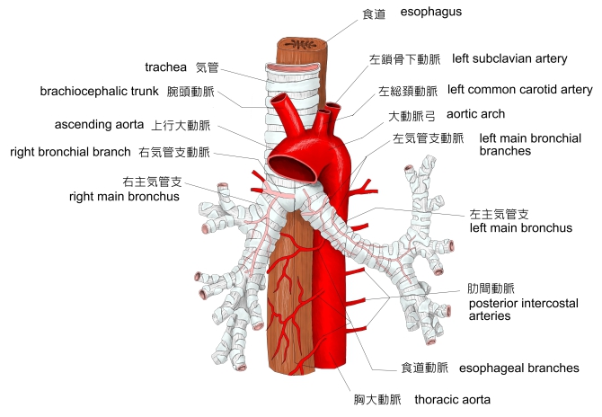
■ 胸 部:thoracic part ■ ・胸腔に入ると次第に気管の左側を走り、気管分岐部の少し下では左気管支のほぼ後ろを通る。 ・胸椎および胸大動脈の前を(はじめ胸大動脈の右側に位置するが次第にその前に出る)下行して横隔膜の食道裂孔を通って腹腔へ。(横隔膜を貫くときには大動脈の前左方を走るようになる。) ・上部では、食道の右側を奇静脈が、後側を胸管が上方に向かって走る。
■ 腹 部:abdominal part ■ ・腹腔に入って少し左方(ときに上方)に曲がり、第11胸椎の前左側で胃の噴門に達する。 ・3部のうちで最も短い。
「日本人体解剖学」には以下のような解説文が見られる。
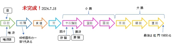
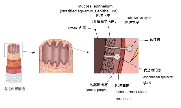
以下に各層の解説を記すが、参考にしたのは「日本人体解剖学」となる。 食道の構造が分かるイラストを掲載しているサイト
【粘 膜】 ■粘膜上皮■ ・重層扁平上皮(stratified squamous epithelium)より成る。 ・一般に角化(keratinization)していないが、場所によっては軽い角化が見られる。 ・上皮細胞の中にはランゲルハンス細胞が存在(免疫的防御に関係) ・胃粘膜上皮との移行部は鋸歯状の境界線が存在
■粘膜固有層■ ・上皮との間に小さな乳頭を形成 (下)食道噴門腺 (esophageal cardiac gland) 常在性にあらず 食道最下部に限局して見られる粘液腺 上食道噴門腺 ※構造的には下食道噴門腺と同じ 食道の上端から第5気管軟骨の高さに約70%の頻度(成人)に出現、胎児では約90%の頻度。
■粘膜筋板■ ・食道の長軸に沿って並ぶ平滑筋線維から成る。 ・消化管の中で最も発育がよく、特に食道下部は上部に比べてよく発達。 ・粘膜筋板は、上方では咽頭の線維膜に、下方では胃の粘膜筋板に移行する。
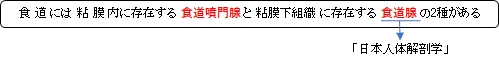
【 粘膜下組織 (submucous tissue) 】 ・ヒダを形成する疎性結合組織、食道腺(混合腺、粘液細胞優先)が見られるが、その発達は個人差が大きい。 ・腺の導管はしばしば途中で膨大し、管の周囲にはしばしばリンパ球の集団が見られることが多い。 ・粘膜下神経叢(マイスネルの神経叢)の発達は弱く分かりにくい。
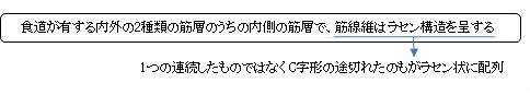
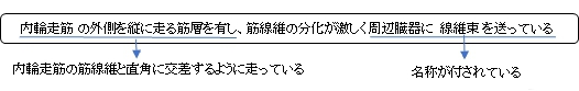
【筋 層】 食道上方1/3部では横紋筋、中1/3では横紋筋と平滑筋、下方1/3ではすべて平滑筋からなる。内輪走筋・外縦走筋に分かれる。 ■ 内輪走筋 ■
・連続したラセン状ではなく、C字形にとぎれたものがラセン状に配列されている。 ・外縦走筋は内輪走筋と直角の方向に走る。 ・食物塊は食道の筋層の蠕動によって運ばれる。
■ 外縦走筋 ■
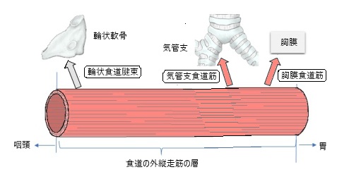
・内輪走筋に比べて著しく分化し、その線維の一部を隣接臓器に送り以下のような呼称が付く。 ※以下、インターネットで色々と調べてみたが参考にできる資料が少なく十分な解説とは言えない。 また、AIなども利用したため、場合によっては正確性に欠ける可能性もある。
1. 輪状食道腱束 (cricoesophageal tendon) 輪状軟骨と食道の間に位置する結合組織の束で、これが食道の運動を支援。輪状食道腱束は、特に嚥下(飲み込み)の際に重要な役割を果たします。 2. 気管支食道筋 (bronchoesophageal muscle) 気管支と食道の間に位置し、両者の運動を調整する役割を果たす。特に呼吸や嚥下のプロセスにおいて重要。 3. 胸膜食道筋 (pleuroesophageal muscle) 胸膜と食道の間に位置し、呼吸と消化の機能をサポートする役割を果た ・内輪走筋と外縦走筋の間には神経細胞をもつ筋層間神経叢(アウエルバッハの神経叢)が発達している。 ・上方の横紋筋部では神経叢の発達は悪い。
【 外 膜 】 食道と隣接臓器または体壁とを結合する厚い線維膜で、比較的太い神経および血管を伴う。
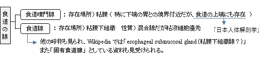
【食道噴門腺:Esophageal cardiac glands】 mucous glands located near the cardiac orifice (esophago-gastric junction) in the lamina propria mucosae. They secrete neutral mucin that protects the esophagus from acidic gastric juices. They are simple tubular or branched tubular glands. 【語 句】 ・cardiac orifice:噴門 ・lamina propria mucosae:粘膜固有層 ・secret:分泌する ・neutral mucin:中性ムチン ・acidic gastric juices:酸性胃液 ・tubular glands:管状腺
【食道腺】 以下はWikipediaの解説文となる。 Esophageal submucosal glands are compound tubulo-alveolar glands. Some serous cells are present. These glands are more numerous in the upper third of the esophagus. They secrete acid mucin for lubrication. 【語 句】 ・Esophageal submucosal glands:食道粘膜下腺? ・tubulo-alveolar glands:管状胞状腺 ・serous cell:漿液細胞 ・lubrication:潤滑
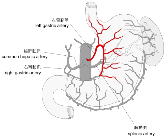
【動 脈】 上部は下甲状腺動脈からの食道枝、中部は食道動脈(胸大動脈の枝)、下部は左胃動脈からの食道枝が分布する。
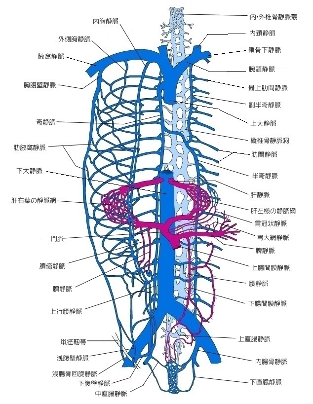
【静 脈】 食道静脈、下甲状腺静脈、奇静脈、半奇静脈、(奇静脈を介して上大静脈と連絡する)、胃冠状静脈(この静脈により門脈と交通する)
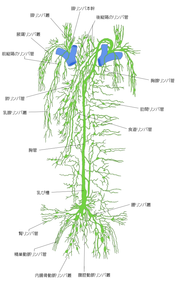
【リンパ管】 食道頚部のものは深頚リンパ節および浅頚リンパ節に、食道胸部のものは後縦隔リンパ節にそそぐ。
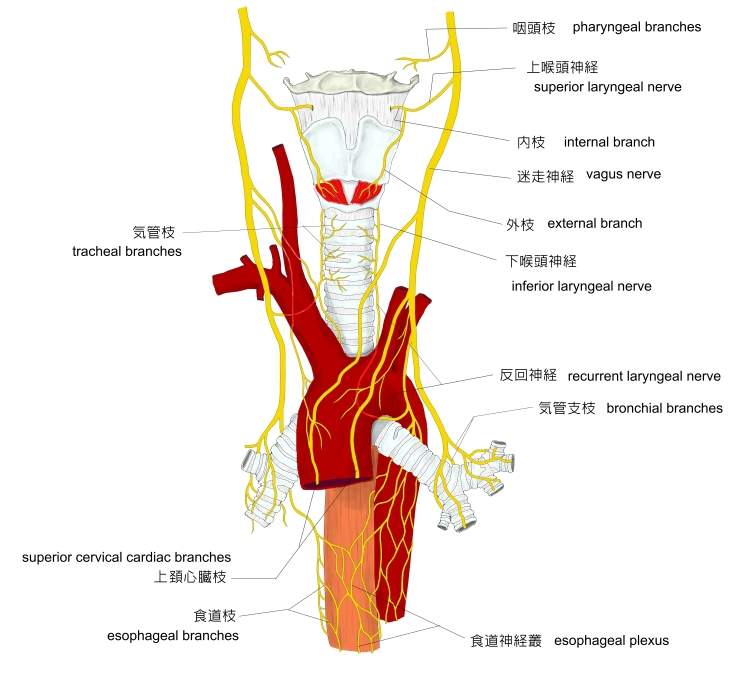
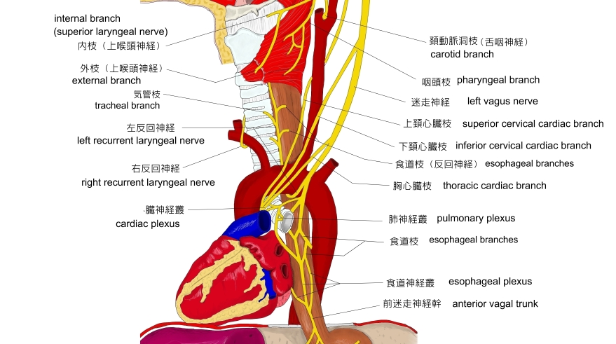
【神 経】 前・後食道神経叢の枝(迷走神経、交感神経幹)。知覚枝は第5~第8胸神経からくる。
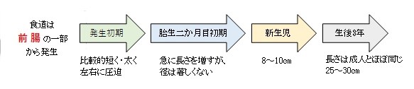
食道は、前腸の一部から発生する。発生の初期には比較的短く、太く、左右から圧迫されている。胎生2か月目の初期になると急に長くなるが、径の発育はあまり著しくない。新生児ではほぼ8~10㎝であるが、生後3年で長さはおよそ、その3倍となる。

以下は「Wikipedia」の解説文となる。 The esophagus (American English) or oesophagus (British English) (/ɪˈsɒfəɡəs/), commonly known as the food pipe or gullet, is an organ in vertebrates through which food passes, aided by peristaltic contractions, from the pharynx to the stomach. The esophagus is a fibromuscular tube, about 25 centimetres long in adults, which travels behind the trachea and heart, passes through the diaphragm and empties into the uppermost region of the stomach. During swallowing, the epiglottis tilts backwards to prevent food from going down the larynx and lungs. The word esophagus is the Greek word οἰσοφάγος oisophagos, meaning "gullet". 【語 句】 ・: ・: ・: ・: ・: ・: ・: ・: ・: ・: ・: ・: ・: ・: ・: ・: ・: ・: ・: ・: ・: ・: ・: ・: ・: ・: ・: ・: ・: ・: ・: ・: ・: ・: ・: ・: ・: ・: ・: ・: ・: ・: ・: ・: ・: ・: ・: ・:
■ 写真やイラストを掲載しているサイト ■
|
||||||||||||||||||||||||||||||||||||||||||||||||||||||||||||||||||||||||||||||||||||||||||
+ |