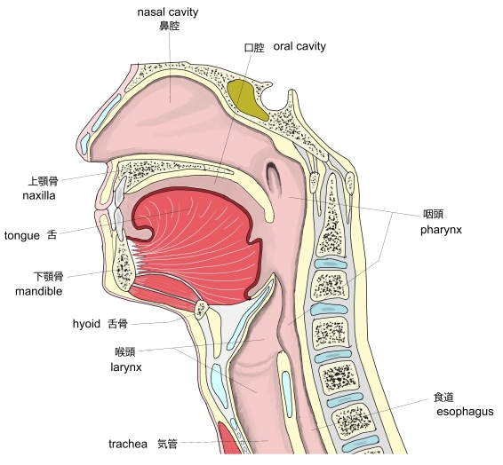
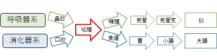
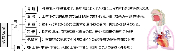
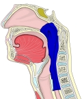
以下は呼吸器系と消化器系を構成する空隙を簡単に表したものとなる。
※付属する器官は省略している。
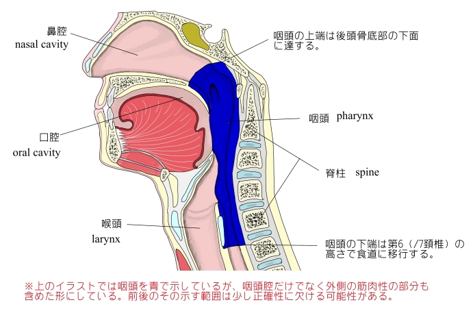
以下は「日本人体解剖学」の解説文となる。
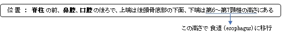
「上端は後頭骨底部の下面に達し、下端は第6~第7頸椎の高さで食道に移行する。その長さは約12㎝で、上方は広く、下方は狭い円錐形を呈する。上方は広く、下方は狭い円錐形を呈する。咽頭は、下方に行くにつれて次第に管状となり、前・後壁は接近し、喉頭の高さでは食道に食塊が通過するときを除いては列隙状である。咽頭内面は粘膜に覆われるが、鼻部の粘膜は多列線毛円柱上皮、口部と喉頭部の粘膜は重層扁平上皮である。」

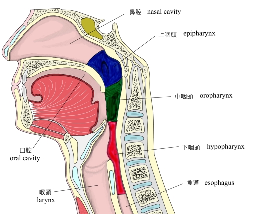
咽頭は以下の3つの部にわけることができる。詳しくはそれぞれの解説ページを参照のこと。
1 |
|
nasal part of pharynx
( epipharynx, nasopharynx ) |
|
2 |
|
oral part of pharynx
(oropharynx, mesopharynx? ) |
3 |
|
laryngeal part of pharynx
( hypopharynx ) |
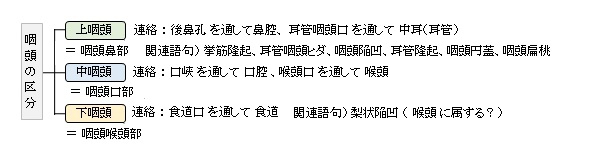
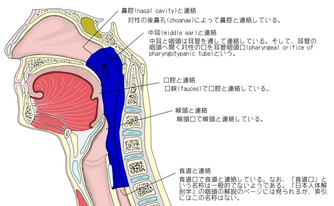
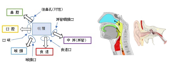
咽頭は「口/孔」で以下のものに連絡をしている。

・鼻腔:後鼻孔(対性)で鼻腔と連絡している。
・口腔:口峡で口腔と連絡している。
・喉頭:喉頭口で喉頭と連絡している。
・食道:食道口で食道と連絡している。
・中耳:耳管咽頭口(対性)で耳管を通じて中耳(鼓室)と連絡する。

■ 粘 膜 ■
咽頭の粘膜は淡紅色を呈する。
1 |
咽頭鼻部の前部 |
多列線毛円柱上皮からなる |
⇒ 呼吸器系の性状 |
2 |
咽頭鼻部の後壁、咽頭口部、および咽頭咽頭部 |
重層扁平上皮からなる |
⇒ 消化器系の性状 |
「粘膜固有層は、線毛上皮の存在する部では乳頭を欠き、重層扁平上皮の部では乳頭を生じる。」
「咽頭円蓋では、リンパ小節がたくさん集まって咽頭扁桃をつくる。」
「咽頭腺は、主として粘液性の小さな混合腺である。すなわち、咽頭円蓋部では混合性、下部に進むにつれて粘液性となり、咽頭口付近に至ると再び混合性となる。」
「粘膜筋板は咽頭粘膜では、まだみられない。」
「粘膜下組織は、頭蓋底の近くの筋層を欠く部では強靭な線維膜からなる。」
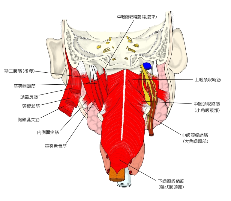
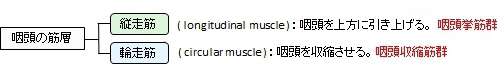
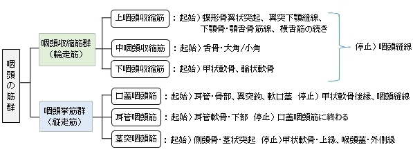
■ 筋 層 ■
咽頭の筋層 (横紋筋) は以下の2群よりなる。
・「筋を欠く最上部には咽頭頭底板がある。」(日本人体解剖学 (下巻) ) )
以下は咽頭の筋群を簡単に表した図となる。
⇒ 咽頭の筋のイラストを掲載しているサイトⅠ
⇒ 咽頭の筋のイラストを掲載しているサイトⅡ
⇒ 咽頭の筋のイラストを掲載しているサイトⅢ
■ 外 膜 ■
弾性繊維に富む疎性結合組織からなる。

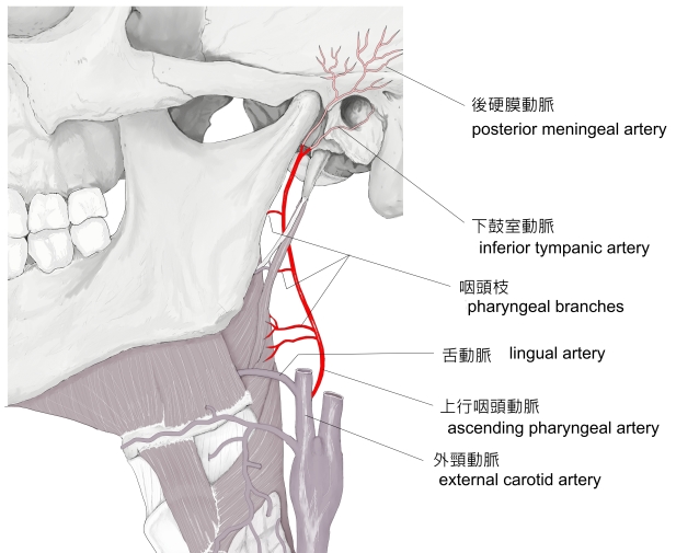
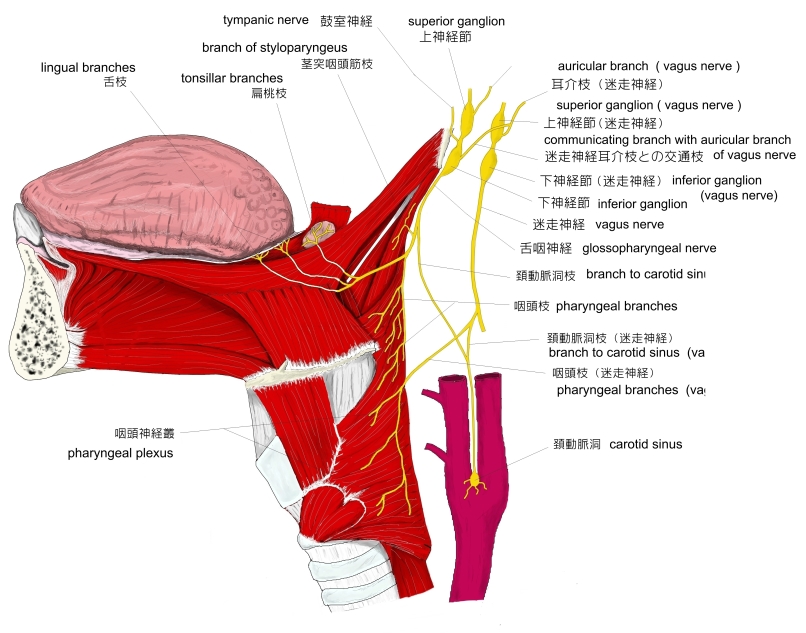
咽頭と関係する脈管や神経は以下になる。


The pharynx (plural: pharynges) is an organ found in vertebrates and invertebrates[citation needed] , though the structure is not universally the same across the species. In humans the pharynx is part of the digestive system and also of the conducting zone of the respiratory system. The conducting zone also includes the nose, larynx, trachea, bronchi, andbronchioles, and their function is to filter, warm, and moisten air and conduct it into the lungs.[citation needed] It makes up the part of the throat situated immediately posterior to the nasal cavity, posterior to the mouth and superior to the esophagus and larynx. The human pharynx is conventionally divided into three sections: the nasopharynx, the oropharynx and the laryngopharynx. It is also important in vocalization.
Structure[edit]
Nasopharynx[edit]
The upper portion of the pharynx, the nasopharynx, extends from the base of the skull to the upper surface of the soft palate.[1] It includes the space between the internal nares and the soft palate and lies above the oral cavity. The adenoids, also known as the pharyngeal tonsils, are lymphoid tissue structures located in the posterior wall of the nasopharynx.
Upper respiratory system, with nasopharynx, oropharynx and laryngopharynx labeled at right.
Polyps or mucus can obstruct the nasopharynx, as can congestion due to an upper respiratory infection. The eustachian tubes, which connect the middle ear to the pharynx, open into the nasopharynx. The opening and closing of the eustachian tubes serves to equalize the barometric pressure in the middle ear with that of the ambient atmosphere.
The anterior aspect of the nasopharynx communicates through the choanae with the nasal cavities. On its lateral walls are the pharyngeal ostia of the auditory tube, somewhat triangular in shape, and bounded behind by a firm prominence, the torus tubarius or cushion, caused by the medial end of the cartilage of the tube that elevates the mucous membrane. Two folds arise from the cartilaginous opening:
Behind the opening of the auditory tube is a deep recess, the pharyngeal recess (also referred to as the fossa of Rosenmüller). On the posterior wall is a prominence, best marked in childhood, produced by a mass of lymphoid tissue, which is known as the pharyngeal tonsil. Superior to the pharyngeal tonsil, in the midline, an irregular flask-shaped depression of the mucous membrane sometimes extends up as far as the basilar process of the occipital bone, this is known as the pharyngeal bursa.
Oropharynx[edit]
The oropharynx lies behind the oral cavity, extending from the uvula to the level of the hyoid bone. It opens anteriorly, through the isthmus faucium, into the mouth, while in its lateral wall, between the Palatoglossal arch and the Palatopharyngeal arch, is the palatine tonsil.[2] The anterior wall consists of the base of the tongue and the epiglottic vallecula; the lateral wall is made up of the tonsil, tonsillar fossa, and tonsillar (faucial) pillars; the superior wall consists of the inferior surface of the soft palate and the uvula. Because both food and air pass through the pharynx, a flap of connective tissue called the epiglottis closes over the glottis when food is swallowed to prevent aspiration. The oropharynx is lined by non-keratinised squamous stratified epithelium.
The HACEK organisms (Haemophilus, Actinobacillus actinomycetemcomitans, Cardiobacterium hominis, Eikenella corrodens, Kingella) are part of the normal oropharyngeal flora, which grow slowly, prefer a carbon dioxide-enriched atmosphere, and share an enhanced capacity to produce endocardial infections, especially in young children.[3] Fusobacterium is a pathogen.[4]
Laryngopharynx[edit]
The laryngopharynx, (Latin: pars laryngea pharyngis), is the caudal part of the pharynx; it is the part of the throat that connects to the esophagus. It lies inferior to the epiglottis and extends to the location where this common pathway diverges into the respiratory (larynx) and digestive (esophagus) pathways. At that point, the laryngopharynx is continuous with the esophagus posteriorly. The esophagus conducts food and fluids to the stomach; air enters the larynx anteriorly. During swallowing, food has the "right of way", and air passage temporarily stops. Corresponding roughly to the area located between the 4th and 6th cervical vertebrae, the superior boundary of the laryngopharynx is at the level of the hyoid bone. The laryngopharynx includes three major sites: the pyriform sinus, postcricoid area, and the posterior pharyngeal wall. Like the oropharynx above it, the laryngopharynx serves as a passageway for food and air and is lined with a stratified squamous epithelium. It is innervated by the pharyngeal plexus.
The vascular supply to the hypopharynx includes the superior thyroid artery, the lingual artery and the ascending pharyngeal artery. The primary neural supply is from both the vagus and glossopharyngeal nerves. The vagus nerve provides a branch termed "Arnolds Nerve" which also supplies the external auditory canal, thus hypophayrngeal cancer can result in referred otalgia. This nerve is also responsible for the ear-cough reflex in which stimulation of the ear canal results in a person coughing.
【 語 句 】
・: ・: ・: ・: ・: ・: ・: ・: ・: ・: ・: ・: ・: ・: ・: ・: ・: ・: ・: ・: ・: ・: ・: ・: ・: ・: ・: ・: ・: ・: ・: ・: ・: ・: ・:

・イラストを掲載しているサイトⅠ
・イラストを掲載しているサイトⅡ
・イラストを掲載しているサイトⅢ
・イラストを掲載しているサイトⅣ
・イラストを掲載しているサイトⅣ

|