
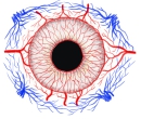
・「血管に富む」(船戸和也のHP)

以下は「Rauber-Kopsh解剖」の解説文となる。
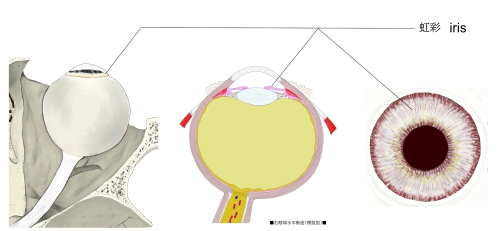
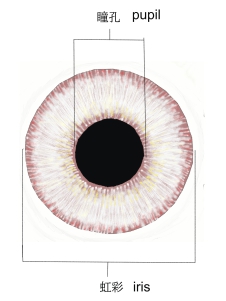
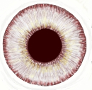
「虹彩の色は個体によって実にさまざまである,金髪の人では青ないし灰色のことが普通で
,緑を帯びていることさえある.褐色または黒色の頭毛の人は虹彩もたいてい暗い色調で,
褐色ないし黒褐色を呈し,虹彩の全体が一様な色のこともあり,霜降りになっていることもある.
(小口昌美(日本医科大学雑誌,8巻3号,1937)によれば日本人の虹彩の色は乳児期には
黒褐色が最も多く,幼児期には濃褐色,学童期から壮年期まで褐色,老年期には褐色および
淡褐色が最も多いという.)青い虹彩では虹彩の結合組織層に色素がない.しかし後面の色素
はどの場合にもある.褐色の虹彩では結合組織性の虹彩支質に,程度の差こそあれ色素が
かなり強く沈着している.白子の虹彩はまったく色素を欠き,多数の血管のために虹彩が赤
くみえ,光線をさえぎる隔膜としての虹彩の使命をごくわずかしか果しえない. 」

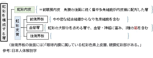
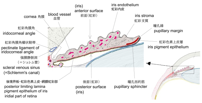
「船戸和也のHP」では虹彩の構成を以下のように解説している。
「虹彩内皮、虹彩支質、虹彩筋、虹彩色素上皮層より構成され、血管に富む。」
|
|
|
|
|
|
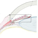
| 虹彩(横断面)
|
|
|
|
|
|

動脈 |
|
|
|
|
小動脈 |
|
静脈 |
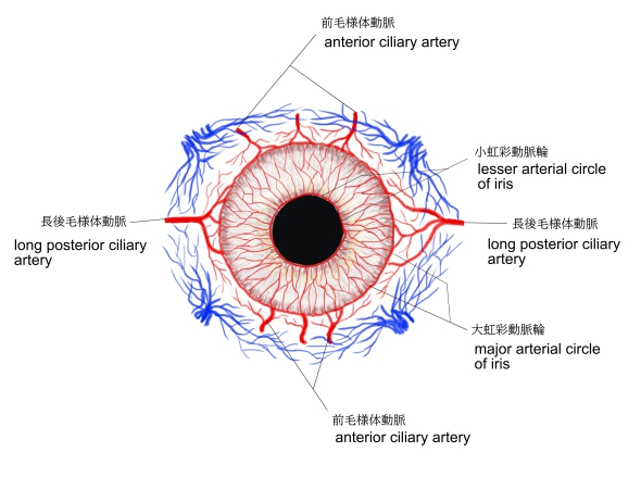
「上記の動脈伴う静脈のほか、渦静脈に流入する。」(船戸和也のHP) |
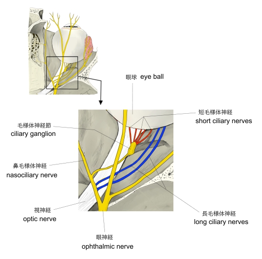
神経 |
|
|
|
|
|
|
|
|
虹彩の動脈 |
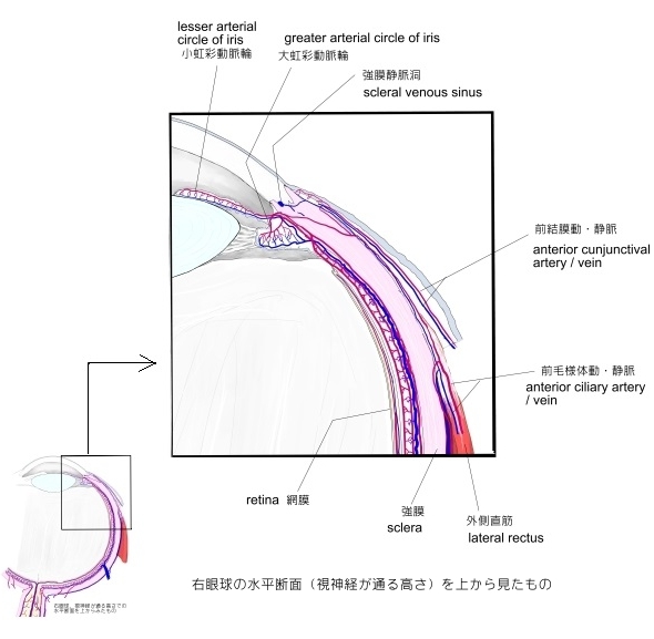
眼球の脈管 |
長・短毛様体神経 |
|

|
|
|
|
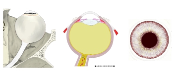
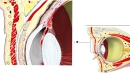
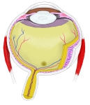
| 眼球(横断面) |
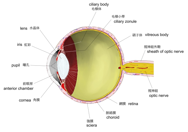
眼球(縦断面) |
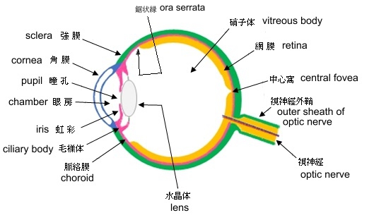
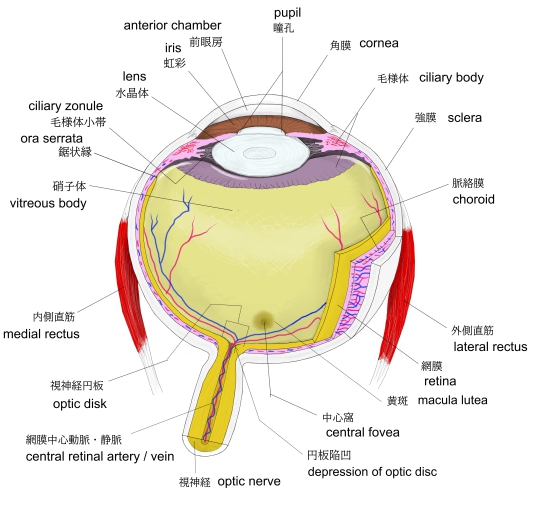
眼球・模型図 |
眼窩・眼球 |
 |
 |
 |
 |
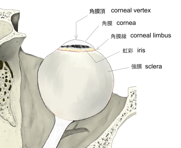
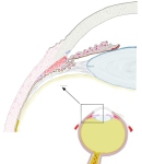
眼球・前面拡大-1 |
眼球・前面拡大-2 |
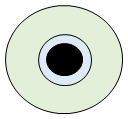
虹彩・模型図(前面) |
虹彩(前面) |
| |
 |
|
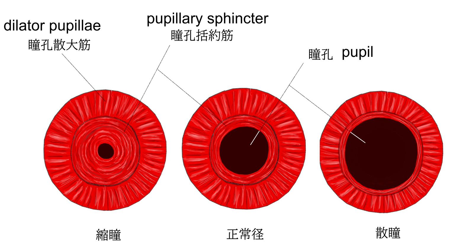
瞳孔の大きさの変化 |
眼球・横断面-1 |
|

か行 |
く |
|
け |
|
こ |
|
さ行 |
し |
|
せ |
|
た行 |
た |
|
と |
|
は行 |
ふ |
|
ま行 |
む |
|
も |
|

以下は「Wikipedia」の解説文となる。
In humans and most mammals and birds, the iris (plural: irides or irises) is a thin, annular structure in the eye, responsible for controlling the diameter and size of the pupil, and thus the amount of light reaching the retina. Eye color is defined by the iris. In optical terms, the pupil is the eye's aperture, while the iris is the diaphragm.
【 語 句 】
・pupil:瞳孔 ・retina:網膜 ・define:定義する ・aperture:穴、隙間 ・diaphragm:隔膜
【Structure】
The iris consists of two layers: the front pigmented fibrovascular layer known as a stroma and, beneath the stroma, pigmented epithelial cells.
The stroma is connected to a sphincter muscle (sphincter pupillae), which contracts the pupil in a circular motion, and a set of dilator muscles (dilator pupillae), which pull the iris radially to enlarge the pupil, pulling it in folds.
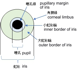
The sphincter pupillae is the opposing muscle of the dilator pupillae. The pupil's diameter, and thus the inner border of the iris, changes size when constricting or dilating. The outer border of the iris does not change size. The constricting muscle is located on the inner border.
【 語 句 】
・pigmented:色素沈着 ・fibrovascular:血管結合組織の ・stroma:支質 ・epithelial cell:上皮細胞 ・sphincter pupillae:瞳孔括約筋 ・contract:縮める ・dilator pupillae:瞳孔散大筋 ・fold:ヒダ ・opposing muscle:対抗筋 ・constrict:収縮させる
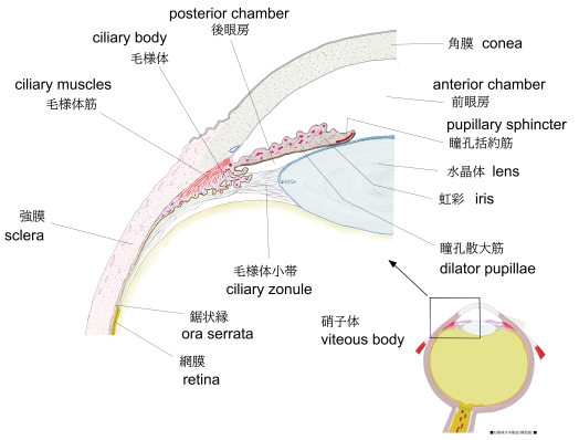
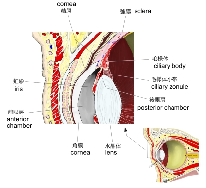
The back surface is covered by a heavily pigmented epithelial layer that is two cells thick (the iris pigment epithelium), but the front surface has no epithelium. This anterior surface projects as the dilator muscles. The high pigment content blocks light from passing through the iris to the retina, restricting it to the pupil. The outer edge of the iris, known as the root, is attached to the sclera and the anterior ciliary body. The iris and ciliary body together are known as the anterior uvea. Just in front of the root of the iris is the region referred to as the trabecular meshwork, through which the aqueous humour constantly drains out of the eye, with the result that diseases of the iris often have important effects on intraocular pressure and indirectly on vision. The iris along with the anterior ciliary body provide a secondary pathway for aqueous humour to drain from the eye.
- The pupillary zone is the inner region whose edge forms the boundary of the pupil.
- The ciliary zone is the rest of the iris that extends to its origin at the ciliary body.
【 語 句 】
・restrict:制限する ・sclera:強膜 ・ciliary body:毛様体 ・uvea:眼球血管膜 ・trabecular meshwork:小柱網 ・aqueous humour:房水 ・intraocular pressure:眼圧 ・pupillary zone:瞳孔領域(=小虹彩輪) ・ciliary zone:毛様体領域 (=大虹彩輪)
The collarette is the thickest region of the iris, separating the pupillary portion from the ciliary portion. The collarette is a vestige of the coating of the embryonic pupil. It is typically defined as the region where the sphincter muscle and dilator muscle overlap. Radial ridges extend from the periphery to the pupillary zone, to supply the iris with blood vessels. The root of the iris is the thinnest and most peripheral.
The muscle cells of the iris are smooth muscle in mammals and amphibians, but are striated muscle in reptiles (including birds). Many fish have neither, and, as a result, their irises are unable to dilate and contract, so that the pupil always remains of a fixed size.
【 語 句 】
・collarette:捲縮輪 ・vestige:痕跡 ・embryonic:胎生の ・define:定義する ・periphery:末梢 ・smooth muscle:平滑筋 amphibian:両生類 ・striated muscle:横紋筋 ・reptile:爬虫類
【Front】
- The crypts of Fuchs are a series of openings located on either side of the collarette that allow the stroma and deeper iris tissues to be bathed in aqueous humor. Collagen trabeculae that surround the border of the crypts can be seen in blue irises.
- The midway between the collarette and the origin of the iris: These folds result from changes in the surface of the iris as it dilates.
- Crypts on the base of the iris are additional openings that can be observed close to the outermost part of the ciliary portion of the iris.
【 語 句 】
・trabeculae:線維柱帯 ・crypt:窩? ・fold:ヒダ
【Back】
- The radial contraction folds of Schwalbe are a series of very fine radial folds in the pupillary portion of the iris extending from the pupillary margin to the collarette. They are associated with the scalloped appearance of the pupillary ruff.
- The structural folds of Schwalbe are radial folds extending from the border of the ciliary and pupillary zones that are much broader and more widely spaced, continuous with the "valleys" between the ciliary processes.
- Some of the circular contraction folds are a fine series of ridges that run near the pupillary margin and vary in thickness of the iris pigment epithelium; others are in ciliary portion of iris.
【 語 句 】
・radial:放射線状の ・contraction:収縮 ・scallop:ホタテ貝 ・ruff:ひだ襟 ・structural:構造上の ・ciliary process:毛様体突起 ・pupillary margin:瞳孔縁 ・pigment epithelium:色素上皮
From anterior (front) to posterior (back), the layers of the iris are:
【Development】
The stroma and the anterior border layer of the iris are derived from the neural crest, and behind the stroma of the iris, the sphincter pupillae and dilator pupillae muscles, as well as the iris epithelium, develop from optic cup neuroectoderm.
【 語 句 】
・be derived from ~:~に由来する ・neural crest:神経堤 ・optic cup:眼杯 ・neuroectoderm:神経外胚葉
■ 写真やイラストを掲載しているサイト ■
・ イラストや写真を掲載しているサイト-Ⅰ
・ イラストや写真を掲載しているサイト-Ⅱ
・ イラストや写真を掲載しているサイト-Ⅲ
・ イラストや写真を掲載しているサイト-Ⅳ
・ イラストや写真を掲載しているサイト-Ⅴ