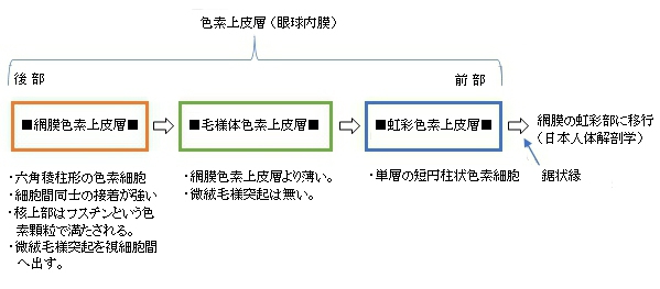
「 日本人体解剖学 」では虹彩を構成する層を以下のように大きく2つに分類し、この虹彩色素上皮層
は含めていない。

それに対して「 船戸和弥のHP 」では以下のように虹彩を構成する層の1つとしている。
「虹彩内皮、虹彩支質、虹彩筋、虹彩色素上皮層より構成され、血管に富む。」

|
|
|
|
|
|
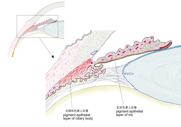
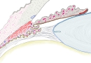
| 毛様体・虹彩色素上皮層
|
|
|
|
|
|
以下は「Wikipedia」の解説文となる。
The iris pigment epithelium (IPE) is a one cell thick layer of cuboidal cells lying behind the iris. The epithelial cells are highly pigmented due to the numerous large melanosomes which pack the cytoplasm of each cell. Towards the central axis, the IPE terminates at the pupillary margin. Peripherally, the IPE is continuous with the inner, non-pigmented layer of the ciliary epithelium. The iris dilator muscle is strictly attached to the anterior side of the iris pigmented epithelium and represents the anterior continuation of the pigmented ciliary epithelium. The ciliary epithelia represent the anterior continuation of the multilayered retina, whose retinal pigmented epithelium (RPE) corresponds to the pigmented ciliary epithelium, while the multilayered sensory retina fades into the non-pigmented ciliary epithelium. Despite their very different functions and histological appearances, these regions have a common origin from the two layers of the embryological optic cup. The melanosomes of the IPE are distinctive, being larger, blacker and rounder than those in the ciliary epithelium or RPE.
【 語 句 】
・pigment:色素 ・melanosomes:メラノソーム ・sytoplasm:原形質、細胞質 ・axis:軸 ・pupillary margin:瞳孔縁 ・peripherally:末梢性に ・ciliary epithelium:毛様体上皮 ・dilator muscle:散大筋 ・strictly:厳密に ・retina:網膜 ・histological: ・embryological:組織学の ・optic cup:眼杯 ・distinctive:特有の
■ 写真やイラストを掲載しているサイト ■
・ イラストや写真を掲載しているサイト-Ⅰ
・ イラストや写真を掲載しているサイト-Ⅱ
・ イラストや写真を掲載しているサイト-Ⅲ
・ イラストや写真を掲載しているサイト-Ⅳ