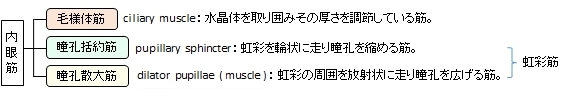
瞳孔散大筋とは

 |
 |
 |
 |
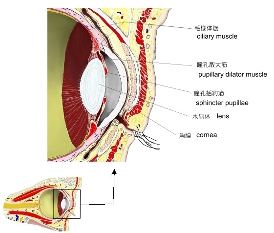
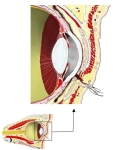
眼窩前部(断面) |
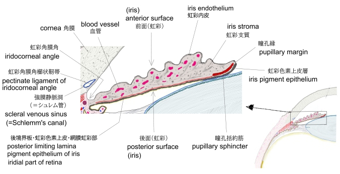
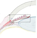
虹彩(横断面)
|
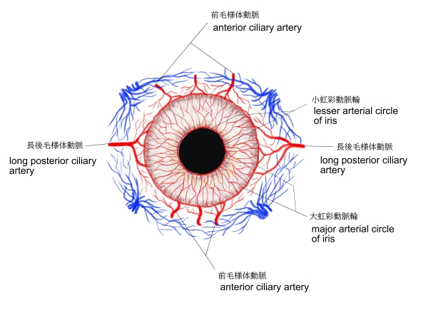
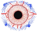
虹彩の血管 |
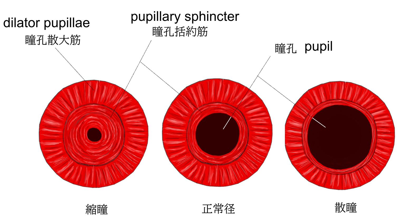
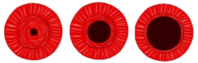
瞳孔の大きさ
|

資料によっては、眼筋を大きく内眼筋と外眼筋に分けているものも見られるが、「 船戸和弥のHP 」や
「 日本人体解剖学 (上巻) 」などでは、この内眼筋を「眼筋」の中に含めていない。その理由は平滑筋だからだと思われる。
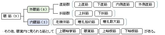
以下は「 日本人体解剖学 」および「 船戸和弥のHP 」でいう「眼筋」で、「 日本人体解剖学 」では一番下の 上眼瞼挙筋 は含めていない。
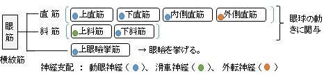
また、以下は眼筋を 外眼筋( 横紋筋 )と 内眼筋( 平滑筋 )に大きく分けた分類となる。
【参考になるサイト】
以下は「Wikipedia」の解説文となる。
「The iris dilator muscle (pupil dilator muscle, pupillary dilator, radial muscle of iris, radiating fibers), is a smooth muscle[2] of the eye, running radially in the iris and therefore fit as a dilator. The pupillary dilator consists of a spokelike arrangement of modified contractile cells called myoepithelial cells. These cells are stimulated by the sympathetic nervous system.[3] When stimulated, the cells contract, widening the pupil and allowing for more light to pass through the eye.
Structure
Innervation:It is innervated by the sympathetic system, which acts by releasing noradrenaline, which acts on α1-receptors.[4] Thus, when presented with a threatening stimuli that activates the fight-or-flight response, this innervation contracts the muscle and dilates the iris, thus temporarily letting more light reach the retina.
The dilator muscle is innervated more specifically by postganglionic sympathetic nerves arising from the superior cervical ganglion as the sympathetic root of ciliary ganglion. From there, they travel via the internal carotid artery through the carotid canal to foramen lacerum. They then enter the middle cranial fossa above foramen lacerum, travel through the cavernous sinus in the middle cranial fossa and then travel with the ophthalmic artery in the optic canal or on the ophthalmic nerve through the superior orbital fissure. From there, they travel with the nasociliary nerve and then the long ciliary nerve. They then pierce the sclera, travel between sclera and choroid to reach the iris dilator muscle. They will also pass through ciliary ganglion and travel in short ciliary nerves to reach the iris dilator muscle.
Function:The pupillary dilator acts to increase the size of the pupil to allow more light to enter the eye. It works in opposition to the pupillary constrictor. Pupil dilation occurs when there is insufficient light for the normal function of the eye, and during heightened sympathetic activity, for example in the "fight or flight reflex."」
■ イラストや写真を掲載しているサイト ■
・イラストや写真を掲載しているサイト-Ⅰ
・イラストや写真を掲載しているサイト-Ⅱ
・イラストや写真を掲載しているサイト-Ⅲ