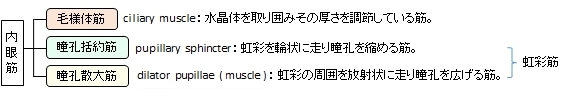
瞳孔括約筋は

 |
 |
 |
 |
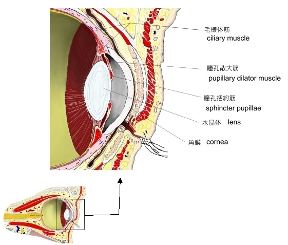
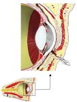
眼窩前部(断面) |
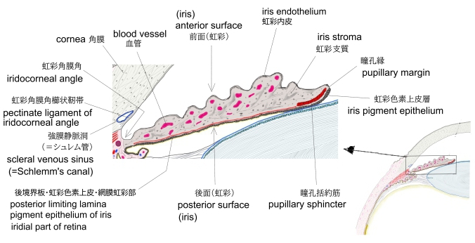
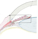
虹彩(横断面)
|
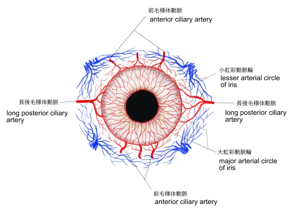
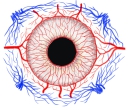
虹彩の血管 |
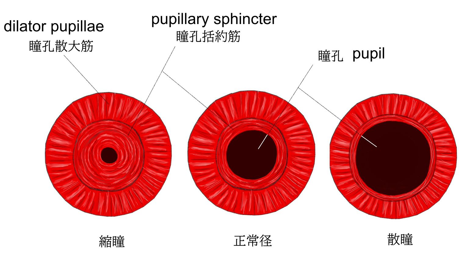
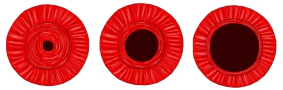
瞳孔の大きさ
|
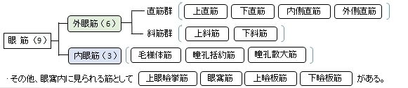
資料によっては、眼筋を大きく内眼筋と外眼筋に分けているものも見られるが、「 船戸和弥のHP 」や
「 日本人体解剖学 (上巻) 」などでは、この内眼筋を「眼筋」の中に含めていない。その理由は平滑筋だからだと思われる。
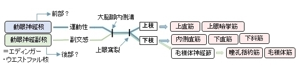
以下は「 日本人体解剖学 」および「 船戸和弥のHP 」でいう「眼筋」で、「 日本人体解剖学 」では一番下の 上眼瞼挙筋 は含めていない。
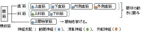
また、以下は眼筋を 外眼筋( 横紋筋 )と 内眼筋( 平滑筋 )に大きく分けた分類となる。
【参考になるサイト】
以下は「Wikipedia」の解説文となる。
「The iris sphincter muscle (pupillary sphincter, pupillary constrictor, circular muscle of iris, circular fibers) is a muscle in the part of the eye called the iris. It encircles the pupil of the iris, appropriate to its function as a constrictor of the pupil.
Comparative Anatomy:It is found in vertebrates and some cephalopods.[citation needed]
General Structure:Initially, all the myocytes are of the smooth muscle type but, later in life, most cells are of the striated muscle type.[2]
Its dimensions are about 0.75 mm wide by 0.15 mm thick.[citation needed]
Mode of Action:In humans, it functions to constrict the pupil in bright light (pupillary light reflex) or during accommodation.[citation needed]
Innervation:It is controlled by parasympathetic fibers that originate from the Edinger-Westphal nucleus, travel along the oculomotor nerve (CN III), synapse in the ciliary ganglion, and then enter the eye via the short ciliary nerves.[citation needed]. The short ciliary nerves then run forward and pierce the sclera at the back of the eye, traveling between the sclera and the choroid to innervate the iris sphincter muscle.」
■ イラストや写真を掲載しているサイト ■
・イラストや写真を掲載しているサイト-Ⅰ
・イラストや写真を掲載しているサイト-Ⅱ
・イラストや写真を掲載しているサイト-Ⅲ