|
|
|
|
|
蝶形骨(上面) |
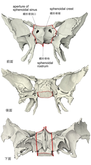
蝶形骨(前・後・下面) |
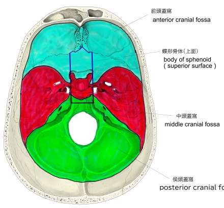
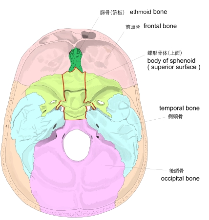
頭蓋窩-1 |
頭蓋窩-2 |
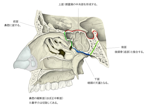
鼻腔(縦断面)
|
「 船戸和弥のHP 」では以下のように解説している。
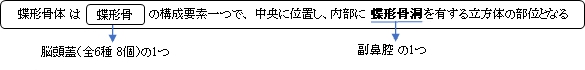
「蝶形骨体は蝶形骨の中央部にあり立方体をなしている。上面中央部には鞍状を呈したトルコ鞍があり、その中央に横位楕円形の下垂体窩がある。トルコ鞍の後方には鞍背という上方に突出した骨板があり、その両側外側端の突起を後床突起という。鞍背の後部は台形をなして後頭骨の底部とともに斜台を形成する。下垂体窩の前には体の前部との境界線である鞍結節とよべる横走する稜があり、その両側端にある中床突起は発育が弱く明瞭なものは少ない。鞍結節の前には細い横走する[視神経]交叉溝があり、その両外側は視神経管につづく。交叉溝の前部は蝶形骨隆起とよばれているが、これは隆起ではなく滑らかな平面である。体の前部は小翼と後部は大翼と結合している。下錐体窩の外側と大翼の根部との間には、内側頚動脈溝という前後に走る溝があり、外側に蝶形骨小舌という突起状の骨板がある。体の下面は鼻腔、咽頭腔の上壁をなし、中央に蝶形骨吻が前下方に突出し鋤骨翼にはさまれる。体の前面中央部には蝶形骨稜という上下に走る稜線があり、篩骨の垂直板と相接する。蝶形骨稜の両側でがいおうに蝶形骨甲介が認められる。これはバルタン小骨ともよばれ、発生学的には篩骨の一部であったものが8~12歳に蝶形骨体と癒合したものでとくに若年頭蓋で著明である。体の内面は空洞状をなし蝶形骨洞とよばれ、その正中部には蝶形骨洞中隔があり、洞を左右に分けている。その前面には蝶形骨洞口という開口部が両側にあり蝶篩陥凹に通じている。」
また、以下は「Wikipedia」の解説文となる。
The body of the sphenoid bone, more or less cubical in shape, is hollowed out in its interior to form two large cavities, the sphenoidal sinuses, which are separated from each other by a septum.
【 語 句 】
・cubical:立方体のように ・sphenoidal sinus:蝶形骨洞 ・septum:隔壁
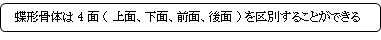
【Superior surface】
The superior surface of the body [Fig. 1] presents in front a prominent spine, the ethmoidal spine, for articulation with the cribriform plate of the ethmoid bone; behind this is a smooth surface slightly raised in the middle line, and grooved on either side for the olfactory lobes of the brain.
This surface is bounded behind by a ridge, which forms the anterior border of a narrow, transverse groove, the prechiasmatic groove, above and behind which lies the optic chiasma; the groove ends on either side in the optic foramen, which transmits the optic nerve and ophthalmic artery into the orbital cavity.
Behind the chiasmatic groove is an elevation, the tuberculum sellae; and behind this is a deep depression, the saddle-shaped sella turcica (Turkish seat), the deepest part of which, the hypophyseal fossa, lodges the pituitary gland.
The anterior boundary of the sella turcica is completed by two small eminences, one on either side, called the middle clinoid processes, while the posterior boundary is formed by a square-shaped plate of bone, the dorsum sellae, ending at its superior angles in two tubercles, the posterior clinoid processes, the size and form of which vary considerably in different individuals.
The posterior clinoid processes deepen the sella turcica, and give attachment to the tentorium cerebelli.
On either side of the dorsum sellae is a notch for the passage of the abducent nerve, and below the notch a sharp process, the petrosal process, which articulates with the apex of the petrous portion of the temporal bone, and forms the medial boundary of the foramen lacerum.
Behind the dorsum sellae is a shallow depression, the clivus, which slopes obliquely backward, and is continuous with the groove on the basilar portion of the occipital bone; it supports the upper part of the pons.
【 語 句 】
・prominent:突出した ・ethmoidal spine:篩骨棘 ・articulate with ~:~と接合する ・cribriform plate:篩板 ・ethmoid bone:篩骨 ・olfactory lobes:嗅脳 ・prechiasmatic groove:視神経交差溝 ・optic chiasma:視交叉 ・optic foramen:視神経孔 ・ophthalmic artery:眼動脈 ・tuberculum sellae:鞍結節 ・sella turcica:トルコ鞍 ・hypophyseal fossa:下垂体窩 ・pituitary gland:下垂体 ・middle clinoid process:中床突起 ・dorsum sellae:鞍背 ・posterior clinoid process:後床突起 ・tentorium cerebelli:小脳テント ・abducent nerve:外転神経 ・petrosal process:岩状突起 ・temporal bone:側頭骨 ・foramen lacerum:破裂孔 ・clivus:斜台 ・occipital bone:後頭骨 ・pons:橋
【Lateral surfaces】
The lateral surfaces of the body are united with the greater wings of the sphenoid and the medial pterygoid plates.
Above the attachment of each greater wing is a broad groove, curved something like the italic letter f; it lodges the internal carotid artery and the cavernous sinus, and is named the carotid sulcus.
Along the posterior part of the lateral margin of this groove, in the angle between the body and greater wing, is a ridge of bone, called the sphenoidal lingula.
【Posterior surfaces】
The posterior surface, quadrilateral in form [Fig. 3], is joined, during infancy and adolescence, to the basilar part of the occipital bone by a plate of cartilage.
Between the eighteenth and twenty-fifth years this becomes ossified, ossification commencing above and extending downward.
【 語 句 】
・greater wings:大翼 ・medial pterygoid plates:内側板 ・internal carotid artery:内頚動脈 ・cavernous sinus:海綿静脈洞 ・carotid sulcus:頚動脈溝 ・ridge:隆起(線) ・sphenoidal lingula:蝶形骨小舌 ・quadrilateral:四辺形の ・infancy:幼少 ・adolescence:思春期 ・cartilage:軟骨 ・ossified:骨化して ・ossification:骨化 ・commence:始める
【Anterior surface】
The anterior surface of the body [Fig. 2] presents, in the middle line, a vertical crest, the sphenoidal crest, which articulates with the perpendicular plate of the ethmoid, and forms part of the nasal septum.
On either side of the crest is an irregular opening leading into the corresponding sphenoidal air sinus.
These sinuses are two large, irregular cavities hollowed out of the interior of the body of the bone, and separated from one another by a bony septum, which is commonly bent to one or the other side.
They vary considerably in form and size, are seldom symmetrical, and are often partially subdivided by irregular bony laminae.
Occasionally, they extend into the basilar part of the occipital bone nearly as far as the foramen magnum. They begin to be developed before birth, and are of a considerable size by the age of six.
【 語 句 】
・sphenoidal crest:蝶形骨稜 ・perpendicular plate:垂直板 ・nasal septum:鼻中隔 ・sphenoidal air sinus:蝶形骨洞? ・hollow:中空の ・laminae:薄板 ・foramen magnum:大後頭孔
They are partially closed, in front and below, by two thin, curved plates of bone, the sphenoidal conchae, leaving in the articulated skull a round opening at the upper part of each sinus by which it communicates with the upper and back part of the nasal cavity and occasionally with the posterior ethmoidal air cells.
The lateral margin of the anterior surface is serrated, and articulates with the lamina papyracea of the ethmoid, completing the posterior ethmoidal cells; the lower margin articulates with the orbital process of the palatine bone, and the upper with the orbital plate of the frontal bone.
【Inferior surface】
The inferior surface presents, in the middle line, a triangular spine, the sphenoidal rostrum, which is continuous with the sphenoidal crest on the anterior surface, and is received in a deep fissure between the alæ of the vomer.
On either side of the rostrum is a projecting lamina, the vaginal process, directed medialward from the base of the medial pterygoid plate, with which it will be described.
【 語 句 】
・sphenoidal conchae:蝶形骨甲介 ・serrated:鋸歯状の ・lamina papyracea:眼窩板 ・posterior ethmoidal air cells:侯篩骨蜂巣 ・orbital process of the palatine bone:篩骨の眼窩突起 ・orbital plate:眼窩板 ・fissure:裂孔 ・alae of the vomer:鋤骨翼 ・vaginal process:鞘状突起 ・medial pterygoid plate:内側板