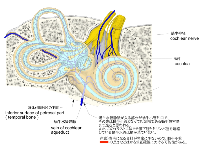


第1回転(=(基)底回転) |
|
第3回転(=尖回転) |
最も小さな回転で、次第に縮小して 蝸牛頂で盲端となって終わる。 |
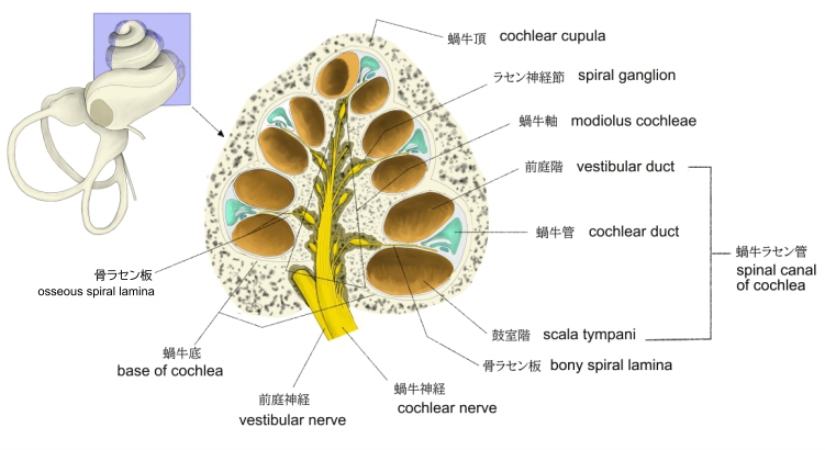
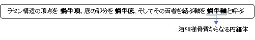
・形状がカタツムリに似ているため「蝸牛」と呼ばれる。
・「中耳側の基部の太さはおよそ 2mm ほどである。」(ウィキペディア)
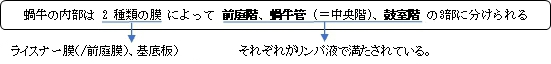
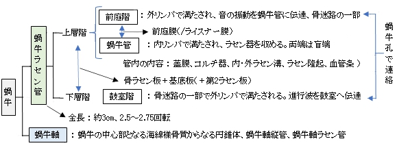
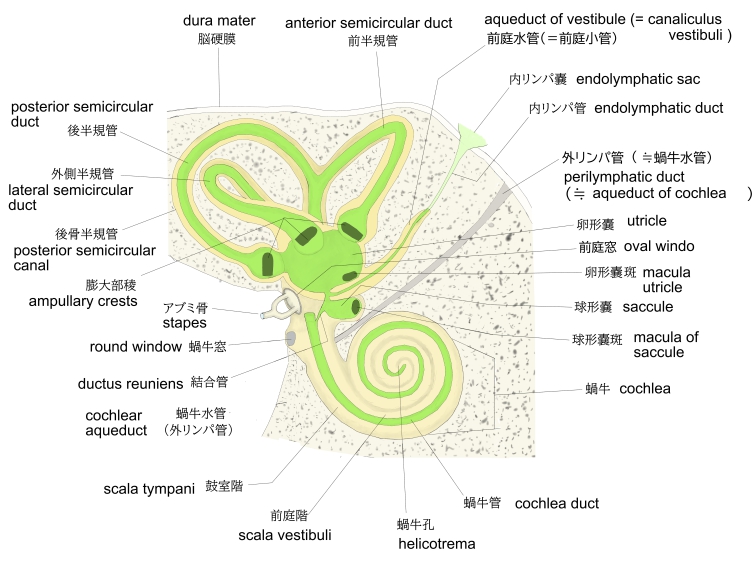
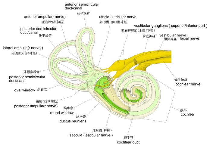
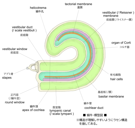
・前庭階と鼓室階は尖端部にある蝸牛孔によってつながっていてる。
・「日本人体解剖学」では蝸牛を蝸牛ラセン管と蝸牛軸の2つに分けて解説している。

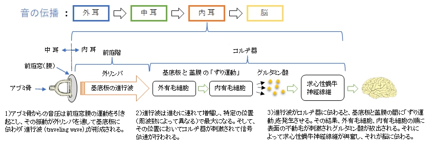
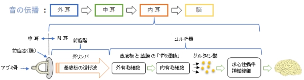
以下「プロメテウス解剖学アトラス(頭部・神経細胞)」を参考にして内耳における「音の伝導」を解説したものになる。
1) アブミ骨からの音圧は前庭窓膜の運動を引き起こす。
2) 前庭窓膜の振動は外リンパを通して基底板(/膜)に伝えられる。
3) 基底板(/膜)に伝わった振動は「進行波(traveling wave)」となって振幅を増加しながら蝸牛頂へと進んでいく。
4) 進行波は特定の位置で(周波数によって異なる)最大となる。
5) その最大となった部位でコルチ器を刺激し信号伝達を行う。
6) 刺激を受けたコルチ器は基底板と蓋膜に最大の歪みを発生させる。
7) その基底板と蓋膜との歪みは「ずり運動」と呼ばれる。
8)「ずり運動」は外有毛細胞の表面に並んでいる不動毛を曲げる。
9)それに反応して、内有毛細胞の不動毛が曲がり、その基底部にグルタミン酸が放出される。
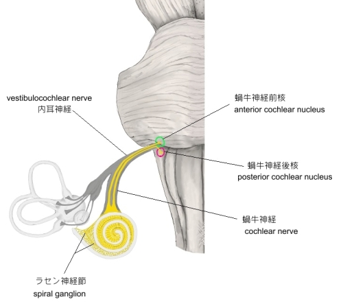
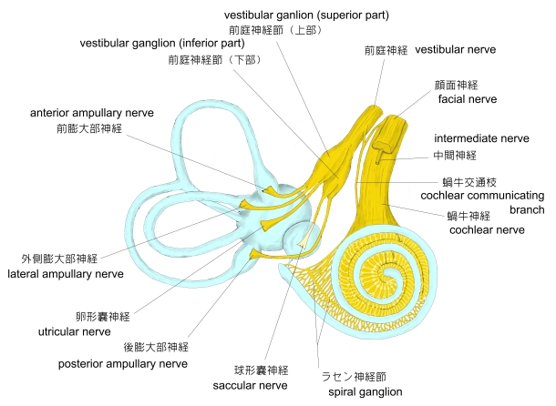
10)放出されたグルタミン酸は求心性蝸牛神経線維を興奮させ、それが脳まで伝わっていく。

1 |
動脈 |
|
2 |
静脈 |
後ラセン状静脈、蝸牛軸ラセン静脈、蝸牛小管静脈 |
3 |
神経 |
|

か行 |
か |
|
き |
|
こ |
|
さ行
|
せ |
|
た行 |
た |
|
ち |
|
ら行 |
ら |
|
以下は「Wikipedia」の解説文となる。
The cochlea is the part of the inner ear involved in hearing. It is a spiral-shaped cavity in the bony labyrinth, in humans making 2.75 turns around its axis, the modiolus.[2][3] A core component of the cochlea is the Organ of Corti, the sensory organ of hearing, which is distributed along the partition separating the fluid chambers in the coiled tapered tube of the cochlea.
The name cochlea derives from Ancient Greek κοχλίας (kokhlias) 'spiral, snail shell'.
【Structure】
Structural diagram of the cochlea showing how fluid pushed in at the oval window moves, deflects the cochlear partition, and bulges back out at the round window.
The cochlea (plural is cochleae) is a spiraled, hollow, conical chamber of bone, in which waves propagate from the base (near the middle ear and the oval window) to the apex (the top or center of the spiral). The spiral canal of the cochlea is a section of the bony labyrinth of the inner ear that is approximately 30 mm long and makes 23⁄4 turns about the modiolus. The cochlear structures include:
【 語 句 】
・: ・: ・: ・: ・: ・: ・: ・: ・: ・: ・: ・: ・: ・: ・: ・: ・: ・: ・: ・: ・: ・: ・: ・: ・: ・: ・: ・: ・: ・: ・: ・: ・: ・: ・: ・:
- Three scalae or chambers:
- the vestibular duct or scala vestibuli (containing perilymph), which lies superior to the cochlear duct and abuts the oval window
- the tympanic duct or scala tympani (containing perilymph), which lies inferior to the cochlear duct and terminates at the round window
- the cochlear duct or scala media (containing endolymph) a region of high potassium ion concentration that the stereocilia of the hair cells project into
- The helicotrema, the location where the tympanic duct and the vestibular duct merge, at the apex of the cochlea
- Reissner's membrane, which separates the vestibular duct from the cochlear duct
- The osseous spiral lamina, a main structural element that separates the cochlear duct from the tympanic duct
- The basilar membrane, a main structural element that separates the cochlear duct from the tympanic duct and determines the mechanical wave propagation properties of the cochlear partition
- The Organ of Corti, the sensory epithelium, a cellular layer on the basilar membrane, in which sensory hair cells are powered by the potential difference between the perilymph and the endolymph
- hair cells, sensory cells in the Organ of Corti, topped with hair-like structures called stereocilia
- The spiral ligament.
The cochlea is a portion of the inner ear that looks like a snail shell (cochlea is Greek for snail).[4] The cochlea receives sound in the form of vibrations, which cause the stereocilia to move. The stereocilia then convert these vibrations into nerve impulses which are taken up to the brain to be interpreted. Two of the three fluid sections are canals and the third is the 'Organ of Corti' which detects pressure impulses that travel along the auditory nerve to the brain. The two canals are called the vestibular canal and the tympanic canal.
【 語 句 】
・: ・: ・: ・: ・: ・: ・: ・: ・: ・: ・: ・: ・: ・: ・: ・: ・: ・: ・: ・: ・: ・: ・: ・: ・: ・: ・: ・: ・: ・: ・: ・: ・: ・: ・: ・:
【Microanatomy】
The walls of the hollow cochlea are made of bone, with a thin, delicate lining of epithelial tissue. This coiled tube is divided through most of its length by an inner membranous partition. Two fluid-filled outer spaces (ducts or scalae) are formed by this dividing membrane. At the top of the snailshell-like coiling tubes, there is a reversal of the direction of the fluid, thus changing the vestibular duct to the tympanic duct. This area is called the helicotrema. This continuation at the helicotrema allows fluid being pushed into the vestibular duct by the oval window to move back out via movement in the tympanic duct and deflection of the round window; since the fluid is nearly incompressible and the bony walls are rigid, it is essential for the conserved fluid volume to exit somewhere.
The lengthwise partition that divides most of the cochlea is itself a fluid-filled tube, the third 'duct'. This central column is called the cochlear duct. Its fluid, endolymph, also contains electrolytes and proteins, but is chemically quite different from perilymph. Whereas the perilymph is rich in sodium ions, the endolymph is rich in potassium ions, which produces an ionic, electrical potential.
【 語 句 】
・: ・: ・: ・: ・: ・: ・: ・: ・: ・: ・: ・: ・: ・: ・: ・: ・: ・: ・: ・: ・: ・: ・: ・: ・: ・: ・: ・: ・: ・: ・: ・: ・: ・: ・: ・:
The hair cells are arranged in four rows in the Organ of Corti along the entire length of the cochlear coil. Three rows consist of outer hair cells (OHCs) and one row consists of inner hair cells (IHCs). The inner hair cells provide the main neural output of the cochlea. The outer hair cells, instead, mainly 'receive' neural input from the brain, which influences their motility as part of the cochlea's mechanical "pre-amplifier". The input to the OHC is from the olivary body via the medial olivocochlear bundle.
The cochlear duct is almost as complex on its own as the ear itself. The cochlear duct is bounded on three sides by the basilar membrane, the stria vascularis, and Reissner's membrane. The stria vascularis is a rich bed of capillaries and secretory cells; Reissner's membrane is a thin membrane that separates endolymph from perilymph; and the basilar membrane is a mechanically somewhat stiff membrane, supporting the receptor organ for hearing, the Organ of Corti, and determines the mechanical wave propagation properties of the cochlear system.
【 語 句 】
・: ・: ・: ・: ・: ・: ・: ・: ・: ・: ・: ・: ・: ・: ・: ・: ・: ・: ・: ・: ・: ・: ・: ・: ・: ・: ・: ・: ・: ・: ・: ・: ・: ・: ・: ・:
【Function】
2:27CC
How sounds make their way from the source to the brain
The cochlea is filled with a watery liquid, the endolymph, which moves in response to the vibrations coming from the middle ear via the oval window. As the fluid moves, the cochlear partition (basilar membrane and organ of Corti) moves; thousands of hair cells sense the motion via their stereocilia, and convert that motion to electrical signals that are communicated via neurotransmitters to many thousands of nerve cells. These primary auditory neurons transform the signals into electrochemical impulses known as action potentials, which travel along the auditory nerve to structures in the brainstem for further processing.
【 語 句 】
・: ・: ・: ・: ・: ・: ・: ・: ・: ・: ・: ・: ・: ・: ・: ・: ・: ・: ・: ・: ・: ・: ・: ・: ・: ・: ・: ・: ・: ・: ・: ・: ・: ・: ・: ・:
■ 写真やイラストを掲載しているサイト ■
・ イラストや写真を掲載しているサイト-Ⅰ
・ イラストや写真を掲載しているサイト-Ⅱ
・ イラストや写真を掲載しているサイト-Ⅲ
・ イラストや写真を掲載しているサイト-Ⅳ
・ イラストや写真を掲載しているサイト-Ⅴ

|