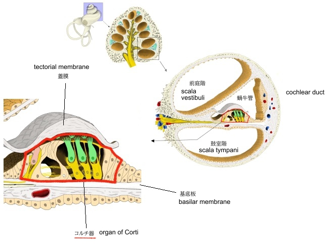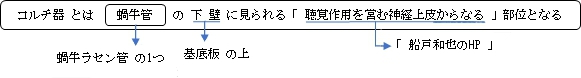
・「ラセン器」とも。
・「その下面は鼓室階被(蓋)層におおわれる。」(日本人体解剖学)
1 |
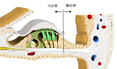
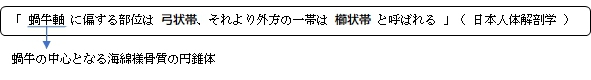
弓状帯 |
・被蓋帯とも ・蝸牛軸に偏する部分で、丈が高く蓋膜で被われている。
・聴覚器にあって最も重要な部位で、有毛細胞と支持細胞よりなる。 |
|
2 |
櫛状帯 |
・弓状帯の外側の部分 ・丈の低い上皮細胞(クラウディウス細胞)で構成される。 |
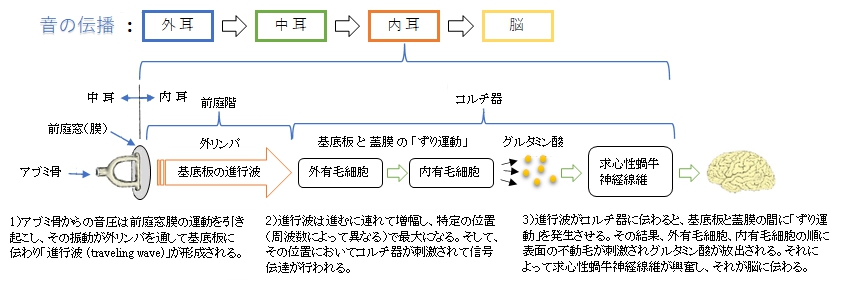
⇒「蝸牛」の「音の伝導」の解説
以下は「新潟大学医学部医学科」のサイトの「内耳のしくみ」の解説文の一部となる。
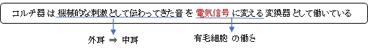
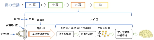
「音入力により、まず、基底板が振動を受け、感覚上皮帯全体が揺れます。 次に、有毛細胞の感覚毛が屈曲し、その頂部にある機械刺激感受性イオンチャネルが開口します。 すると、内リンパ液に豊富にあるK+がチャネルを通って有毛細胞に流入し、細胞を電気興奮させます(図2B)。 すなわち、この過程で、“音の機械的(物理的)刺激が電気信号に変換”されることになります。 結果として、神経伝達物質の放出が発生し、神経線維を通じて脳へと音情報が伝わります。」
|
|
|
|
| コルチ器 |
コルチ器周辺-Ⅰ |
コルチ器周辺-Ⅱ |
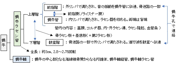
蝸牛管 |
 |
|
|
|
蝸牛ラセン管 |
|
|
|

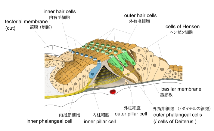
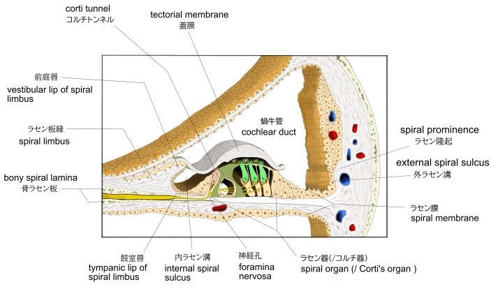
コルチ器を構成する細胞は、大きく有毛細胞と支持細胞に分けることができる。
1 |
有毛細胞 |
a |
|
inner hair cell
・内柱細胞の内側(蝸牛軸側)に一列に並んでいる。
・数は約3500(と言われている)
・自由面には約120本の聴毛を有する。
|
b |
|
outer hair cell
・外柱細胞の外側に複数列(3列~5列)1つおきに並んでいる。
・形は内有毛細胞に似るが、数は4倍近くになる。
・上端は網状膜のそれぞれの孔にはまって固定される。
|
2 |
支持細胞 |
a |
|
|
b |
|
|
c |
|
inner phalangeal cell
・内柱細胞の内側に位置して内有毛細胞を支えている。
|
d |
|
|
e |
|
cell of Hensen
・外指節細胞の外側に位置する複数列の細胞で、コルチ器の傾斜部分を形成している。
|

以下は「Wikipedia」の解説文となる。
The organ of Corti, or spiral organ, is the receptor organ for hearing and is located in the mammalian cochlea. This highly varied strip of epithelial cells allows for transduction of auditory signals into nerve impulses' action potential. Transduction occurs through vibrations of structures in the inner ear causing displacement of cochlear fluid and movement of hair cells at the organ of Corti to produce electrochemical signals.
Italian anatomist Alfonso Giacomo Gaspare Corti (1822–1876) discovered the organ of Corti in 1851. The structure evolved from the basilar papilla and is crucial for mechanotransduction in mammals.
【 語 句 】
・mammalian:哺乳類の ・cochlea:蝸牛 ・epithelial cell:上皮細胞 ・transduction:伝達、変換 ・nerve impulse:神経インパルス ・action potential:活動電位 ・hair cell:有毛細胞 ・basilar papilla:乳頭基部 ・mechanotransduction:機械的シグナル伝達
【Structure】
The organ of Corti is located in the scala media of the cochlea of the inner ear between the vestibular duct and the tympanic duct and is composed of mechanosensory cells, known as hair cells. Strategically positioned on the basilar membrane of the organ of Corti are three rows of outer hair cells (OHCs) and one row of inner hair cells (IHCs).Surrounding these hair cells are supporting cells: Deiters cells, also called phalangeal cells, which have a close relation with the OHCs, and pillar cells, which separate and support both the OHCs and the IHCs.
Projecting from the tops of the hair cells are tiny finger-like projections called stereocilia, which are arranged in a graduated fashion with the shortest stereocilia on the outer rows and the longest in the center. This gradation is thought to be the most important anatomic feature of the organ of Corti because this allows the sensory cells superior tuning capability.
If the cochlea were uncoiled, it would roll out to be about 33 mm long in women and 34 mm in men, with about 2.28 mm of standard deviation for the population. The cochlea is also tonotopically organized, meaning that different frequencies of sound waves interact with different locations on the structure. The base of the cochlea, closest to the outer ear, is the most stiff and narrow and is where the high-frequency sounds are transduced. The apex, or top, of the cochlea is wider and much more flexible and loose and functions as the transduction site for low-frequency sounds.
【 語 句 】
・scala media:中心階 ・inner ear:内耳 ・vestibular duct:前庭階 ・tympanic duct:鼓室階 ・mechanosensory cell:感覚細胞? ・basilar membrane:基底板 ・outer hair cell:外有毛細胞 ・inner hair cell:内有毛細胞 ・Deiters cell:ダイテルス細胞 ・phalangeal cell:指節細胞 ・pillar cell:柱細胞 ・stereocilia:不動毛 ・in a graduated fashion:段階的に ・standard deviation:標準偏差 ・tonotopic:周波数特定の ・interact:相互に作用する
【Function】
The function of the organ of Corti is to convert (transduce) sounds into electrical signals that can be transmitted to the brainstem through the auditory nerve.It is the auricle and middle ear that act as mechanical transformers and amplifiers so that the sound waves end up with amplitudes 22 times greater than when they entered the ear.
■Auditory transduction■
In normal hearing, the majority of the auditory signals that reach the organ of Corti in the first place come from the outer ear. Sound waves enter through the auditory canal and vibrate the tympanic membrane, also known as the eardrum, which vibrates three small bones called the ossicles. As a result, the attached oval window moves and causes movement of the round window, which leads to displacement of the cochlear fluid. However, the stimulation can happen also via direct vibration of the cochlea from the skull. The latter is referred to as Bone Conduction (or BC) hearing, as complementary to the first one described, which is instead called Air Conduction (or AC) hearing. Both AC and BC stimulate the basilar membrane in the same way (Békésy, G.v., Experiments in Hearing. 1960).
The basilar membrane on the tympanic duct presses against the hair cells of the organ as perilymphatic pressure waves pass. The stereocilia atop the IHCs move with this fluid displacement and in response their cation, or positive ion selective, channels are pulled open by cadherin structures called tip links that connect adjacent stereocilia. The organ of Corti, surrounded in potassium-rich fluid endolymph, lies on the basilar membrane at the base of the scala media. Under the organ of Corti is the scala tympani and above it, the scala vestibuli. Both structures exist in a low potassium fluid called perilymph. Because those stereocilia are in the midst of a high concentration of potassium, once their cation channels are pulled open, potassium ions as well as calcium ions flow into the top of the hair cell. With this influx of positive ions the IHC becomes depolarized, opening voltage-gated calcium channels at the basolateral region of the hair cells and triggering the release of the neurotransmitter glutamate. An electrical signal is then sent through the auditory nerve and into the auditory cortex of the brain as a neural message.
【 語 句 】
・auditory nerve:蝸牛神経 ・auricle:外耳 ・middle ear:中耳 ・amplifier:増幅器 ・auditory canal:耳道 ・tympanic membrane:鼓膜 ・ossicles:耳小骨 ・oval window:卵円窓 ・round window:正円窓、蝸牛窓 ・Bone Conduction:骨伝導 ・complementary:補足的な ・Air Conduction:空気伝導 ・basilar membrane:基底板 ・perilymphatic:外リンパの ・atop ~:~の頂上に ・cation:陽イオン ・cadherin:カドヘリン(糖タンパク質の一群) ・adjacent:隣接した ・potassium:カリウム ・endolymph:内リンパ ・scala tympani:鼓室階 ・scala vestibuli:前庭階 ・midst:中央 ・influx:中央 ・depolarize:脱分極させる ・voltage-gated calcium channels:電位開口型カルシウムチャネル ・basolateral:基底外側部の ・neurotransmitter:神経伝達物質 ・glutamate:グルタミン酸 ・auditory cortex:聴覚皮質
■Cochlear amplification■
Further information: Cochlear amplifier
The organ of Corti is also capable of modulating the auditory signal. The outer hair cells (OHCs) can amplify the signal through a process called electromotility where they increase movement of the basilar and tectorial membranes and therefore increase deflection of stereocilia in the IHCs.
A crucial piece to this cochlear amplification is the motor protein prestin, which changes shape based on the voltage potential inside of the hair cell. When the cell is depolarized, prestin shortens, and because it is located on the membrane of OHCs it then pulls on the basilar membrane and increasing how much the membrane is deflected, creating a more intense effect on the inner hair cells (IHCs). When the cell hyperpolarizes prestin lengthens and eases tension on the IHCs, which decreases the neural impulses to the brain. In this way, the hair cell itself is able to modify the auditory signal before it even reaches the brain.
【 語 句 】
・modulate:振幅・周波数などを変調する ・electromotility:電気運動性? ・tectorial membrane:蓋膜 ・deflection:ゆがみ ・crucial:決定的な ・motor protein:モータータンパク質 ・prestin:プレスチン(膜タンパク質) ・hyperpolarize:過分極させる ・modify:修正する
【Development】
The organ of Corti, in between the scala tympani and the scala media, develops after the formation and growth of the cochlear duct. The inner and outer hair cells then differentiate into their appropriate positions and are followed by the organization of the supporting cells. The topology of the supporting cells lends itself to the actual mechanical properties that are needed for the highly specialized sound-induced movements within the organ of Corti.
Development and growth of the organ of Corti relies on specific genes, many of which have been identified in previous research (SOX2, GATA3, EYA1, FOXG1, BMP4, RAC1, and more), to undergo such differentiation. Specifically, the cochlear duct growth and the formation of hair cells within the organ of Corti.
Mutations in the genes expressed in or near the organ of Corti before the differentiation of hair cells will result in a disruption in the differentiation, and potential malfunction of, the organ of Corti.
【 語 句 】
・cochlear duct: ・differentiate:異なってくる ・topology:形態 ・lend:役立つ、適している ・mechanical property:機械的特性 ・mutation:変化、突然変異 ・disruption:崩壊、分裂 ・malfunction:機能不全
■ 写真やイラストを掲載しているサイト ■
・ イラストや写真を掲載しているサイト-Ⅰ
・ イラストや写真を掲載しているサイト-Ⅱ
・ イラストや写真を掲載しているサイト-Ⅲ
・ イラストや写真を掲載しているサイト-Ⅳ
・ イラストや写真を掲載しているサイト-Ⅴ