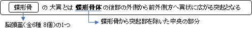

「前外側方へ翼状に広がって中頭蓋窩の一部となり、その前端は尖って少し上向きとなり、頭蓋の側壁の一部となる。」(日本人体解剖学)
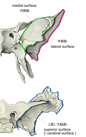
【 面 】
以下に各面の特徴を簡単に記す。(参考:「船戸和也のHP」)
|
名 称 |
特 徴 |
1 |
|
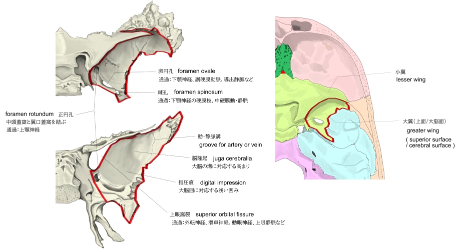
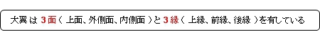
・大脳に面し凹面で、中頭蓋窩の一部を成している。
【認められるもの】
指圧痕、脳隆起、動・静脈溝、孔(正円孔、卵円孔、棘孔)
|
2 |
外側面 |
|
3 |
内側面 |
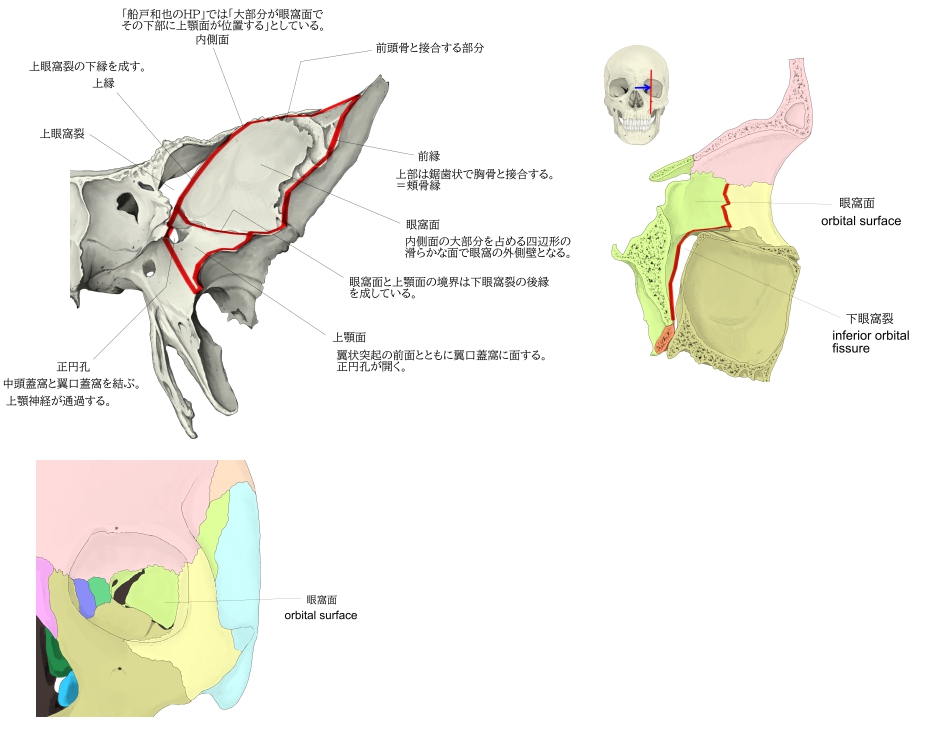
・菱形で眼窩外側壁の形成に預かるため眼窩面といわれる。
・下方にある面を上顎面といい、翼状突起の前面とともに翼口蓋窩に面し、ここに正円孔が開口する。
・「眼窩面と上顎面の境は下眼窩裂の後縁をなす。」(船戸和也のHP)
|
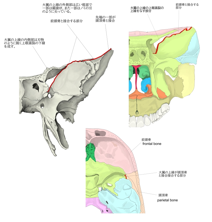
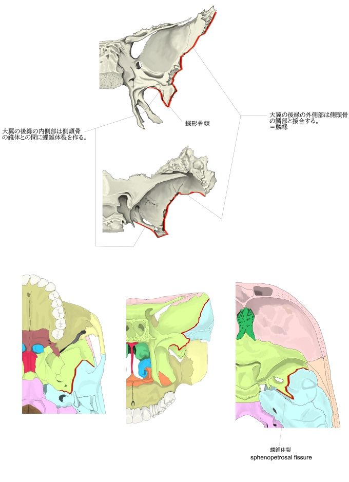
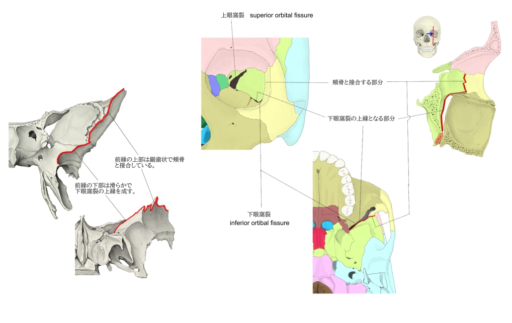
【 縁 】
以下に各縁の特徴を簡単に記す。(参考:「船戸和也のHP」)
|
名 称 |
特 徴 |
1 |
上 縁 |
・大翼の基部から外上方の一番高い先端まで伸びている。=前頭縁
内側:刃物のように鋭く上眼窩裂の下縁を成す。
外側: 広い粗面で、一部は鋸歯状、また一部はノミの刃のように尖っていて、前部で前頭骨、後部で頭頂骨と接合する。
|
2 |
前 縁 |
・上方:鋸歯状で頬骨と接合する。=頬骨縁
・下方:滑らかで下眼窩裂の上縁を成している。
|
3 |
後 縁 |
・外側部(前部):側頭骨の鱗部と接合するため鱗縁と呼ばれる。
・内側部(後部):側頭骨錐体との間に蝶錐体裂をつくる。
蝶形骨棘:上記の両部が合するところの下部へ走る突起
|
以下は「Wikipedia」の解説文となる。
The greater wing of the sphenoid bone, or alisphenoid, is a bony process of the sphenoid bone; there is one on each side, extending from the side of the body of the sphenoid and curving upward, laterally, and backward.
【Structure】
The greater wings of the sphenoid are two strong processes of bone, which arise from the sides of the body, and are curved upward, laterally, and backward; the posterior part of each projects as a triangular process that fits into the angle between the squamous and the petrous part of the temporal bone and presents at its apex a downward-directed process, the spine of sphenoid bone.
【 語 句 】
・body of the sphenoid:蝶形骨体 ・squamous (part) :鱗部 ・petrous part:錐体部 ・temporal bone:側頭部 ・spine of sphenoid bone:蝶形骨棘 ・:
■Cerebral surface■
The superior or cerebral surface of each greater wing forms part of the middle cranial fossa; it is deeply concave, and presents depressions for the convolutions of the temporal lobe of the brain. It has a number of foramina (holes) in it:
【 語 句 】
・middle cranial fossa:中頭蓋窩 ・concave:凹状の ・convolution:脳回 ・temporal lobe:側頭葉 ・foramina:孔 ・foramen rotundum:正円孔 ・aperture:裂孔 ・maxillary nerve:上顎神経 ・foramen ovale:卵円孔 ・mandibular nerve:下顎神経 ・accessory meningeal artery:副硬膜動脈 ・lesser petrosal nerve:小錐体神経 ・sphenoidal emissary foramen:蝶形導出静脈孔 ・pterygoid process:翼状突起 ・scaphoid fossa:舟状窩 ・cavernous sinus:海綿静脈洞 ・foramen spinous:棘孔 ・middle meningeal vessels:中硬膜動・静脈 ・current branch: ・foramen petrosum:錐体孔 ・foramen spinosum:棘孔
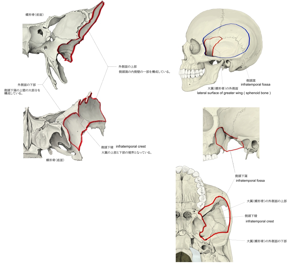
■Lateral surface ■
The lateral surface is convex, and divided by a transverse ridge, the infratemporal crest, into two portions.
- The superior temporal surface, convex from above downward, concave from before backward, forms a part of the temporal fossa, and gives attachment to the temporalis;
- the inferior infratemporal surface, smaller in size and concave, enters into the formation of the infratemporal fossa, and, together with the infratemporal crest, serves as an attachment to the lateral pterygoid muscle.
It is pierced by the foramen ovale and foramen spinosum, and at its posterior part is the sphenoidal spine, which is frequently grooved on its medial surface for the chorda tympani nerve.
To the sphenoidal spine are attached the sphenomandibular ligament and the tensor veli palatini muscle.
Medial to the anterior extremity of the infratemporal crest is a triangular process that serves to increase the attachment of the lateral pterygoid muscle; extending downward and medialward from this process on to the front part of the lateral pterygoid plate is a ridge that forms the anterior limit of the infratemporal surface, and, in the articulated skull, the posterior boundary of the pterygomaxillary fissure.
【 語 句 】
・ridge:隆起(線) ・infratemporal crest:側頭下稜 ・temporal fossa:側頭窩 ・temporalis:側頭筋 ・infratemporal fossa:側頭下窩 ・lateral pterygoid muscle:外側翼突筋 ・chorda tympani nerve:鼓索神経 ・sphenomandibular ligament:蝶下顎靭帯 ・tensor veli palatine muscle:口蓋帆張筋 ・pterygomaxillary fissure:翼上顎裂
■Orbital surface■
The orbital surface of the great wing , smooth, and quadrilateral in shape, is directed forward and medially and forms the posterior part of the lateral wall of the orbit.
- Its upper serrated edge articulates with the orbital plate of the frontal bone.
- Its inferior rounded border forms the postero-lateral boundary of the inferior orbital fissure.
- Its medial sharp margin forms the lower boundary of the superior orbital fissure and has projecting from about its center a little tubercle that gives attachment to the inferior head of the lateral rectus muscle; at the upper part of this margin is a notch for the transmission of a recurrent branch of the lacrimal artery.
- Its lateral margin is serrated and articulates with the zygomatic bone.
- Below the medial end of the superior orbital fissure is a grooved surface, which forms the posterior wall of the pterygopalatine fossa, and is pierced by the foramen rotundum.
【 語 句 】
・quadrilateral: ・orbit: ・serrated: ・articulate with~:~と接合する ・inferior orbital fissure:下眼窩裂 ・superior orbital fissure:上眼窩裂 ・tubercle:小結節 ・lateral rectus muscle:外側直筋 ・notch:切痕 ・lacrimal artery:涙腺動脈 ・zygomatic bone:頬骨 ・pterygopalatine fossa:翼口蓋窩 ・pierce:貫通する ・foramen rotundum:正円孔
【Margin】
Commencing from behind, that portion of the circumference of the great wing that extends from the body to the spine is irregular.
- Its medial half forms the anterior boundary of the foramen lacerum, and presents the posterior aperture of the pterygoid canal for the passage of the corresponding nerve and artery.
- Its lateral half articulates, by means of a synchondrosis, with the petrous portion of the temporal, and between the two bones on the under surface of the skull, is a furrow, the sulcus of the auditory tube, for the lodgement of the cartilaginous part of the auditory tube.
In front of the spine the circumference presents a concave, serrated edge, bevelled at the expense of the inner table below, and of the outer table above, for articulation with the squamous part of the temporal bone.
At the tip of the great wing is a triangular portion, bevelled at the expense of the internal surface, for articulation with the sphenoidal angle of the parietal bone; this region is named the pterion.
Medial to this is a triangular, serrated surface, for articulation with the frontal bone; this surface is continuous medially with the sharp edge that forms the lower boundary of the superior orbital fissure, and laterally with the serrated margin for articulation with the zygomatic bone.
【Development】
The greater wing of the sphenoid bone starts as a separate bone, and is still separate at birth in humans.
【 語 句 】
・commence:始まる ・circumference:周囲 ・foramen lacerum:破裂孔 ・pterygoid canal:翼突管 ・synchondrosis:軟骨結合 ・petrous protion of the temporal:側頭骨の錐体部 ・furrow:溝 ・sulcus of the auditory tube:耳管溝 ・cartilaginous:軟骨性の ・bevelled:傾斜がつけられた? ・at the expense of ~:~を犠牲にして ・articulation with~:~との結合 ・squamous part:鱗部 ・parietal bone:頭頂骨 ・pterion:プテリオン
■ 写真やイラストを掲載しているサイト ■
・ イラストや写真を掲載しているサイト-Ⅰ
・ イラストや写真を掲載しているサイト-Ⅱ
・ イラストや写真を掲載しているサイト-Ⅲ
・ イラストや写真を掲載しているサイト-Ⅳ
・ イラストや写真を掲載しているサイト-Ⅴ