
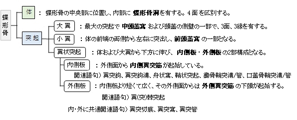
【その他の特徴】
以下に翼状突起の特徴を簡単にまとめてみた。
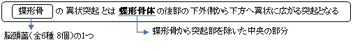
・内側板の根部から内側法に走っている板状の突起を鞘状突起という。
・鞘状突起は鋤骨翼と相対して蝶形骨下面との間に鋤骨鞘突管を形成する。
・鞘状突起の下側を前後に走る管を口蓋骨鞘突管と呼ぶが、しばしば溝状(口蓋骨鞘突溝)となることがある。
(管状、溝状のいずれにしても、蝶口蓋動脈の枝および翼口蓋神経節の枝である外側上後鼻枝が通っている。)
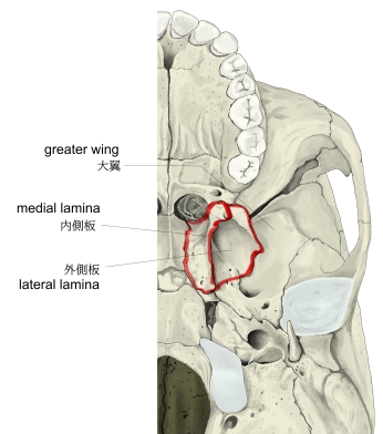
・翼状突起の後面で、内側板および外側板の間にできる深い窩を翼突窩といい、内側翼突筋の起始部となっている。
・翼突窩の上内側方には浅い楕円形の陥凹があり、これを舟状窩といい、口蓋帆張筋の起始部となっている。
・翼状突起の根部を矢状に走る管を翼突管といい、翼突管神経および翼突管動・静脈が通る。
・翼突管の前部の開口は下方に走って溝状となる。これを大口蓋溝といい、上顎骨および口蓋骨と接合して
大口蓋管を形成する。
|
|
|
|
|
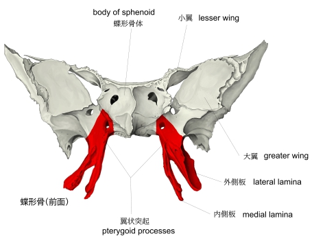
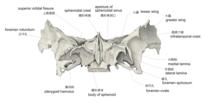
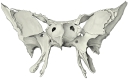
蝶形骨(前面) |
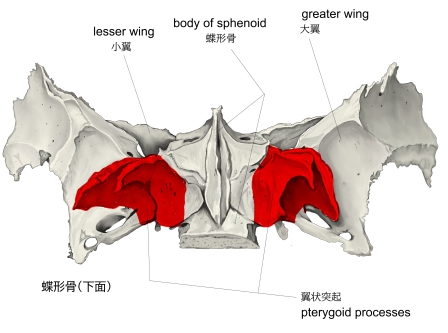
蝶形骨(底面) |
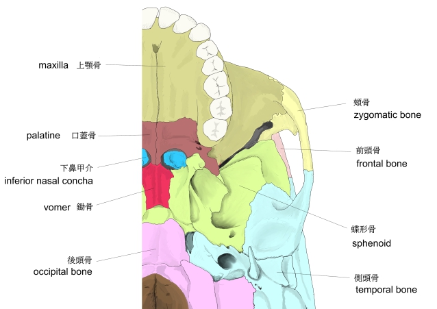
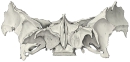
外頭蓋底 |
外頭蓋底 |
|
 |
 |
 |
|
|
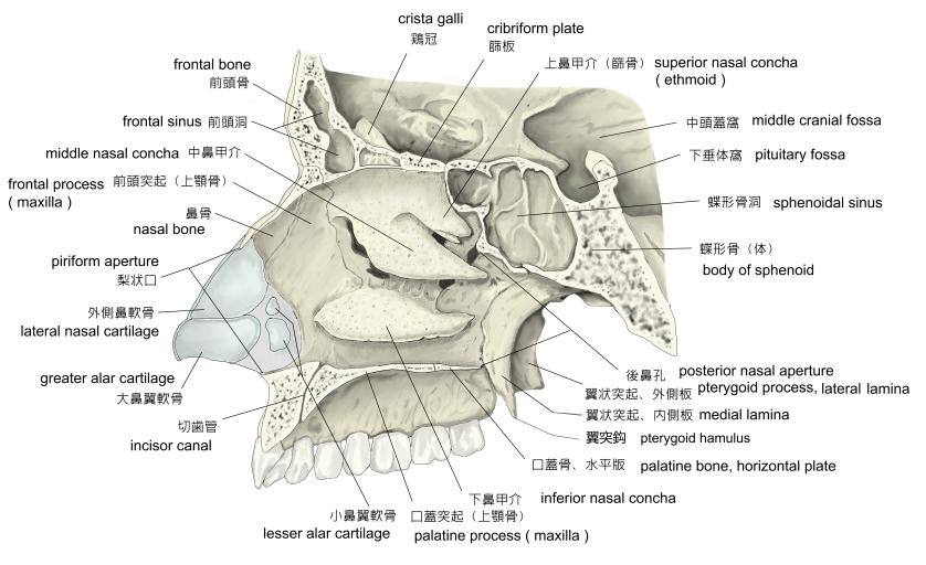
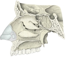
右鼻腔(外側壁) |
蝶形骨(前面) |
蝶形骨(底面) |
|
|
以下は「Wikipedia」の解説文となる。
The pterygoid processes of the sphenoid (from Greek pteryx, pterygos, "wing"), one on either side, descend perpendicularly from the regions where the body and the greater wings of the sphenoid bone unite.
Each process consists of a medial pterygoid plate and a lateral pterygoid plate, the latter of which serve as the origins of the medial and lateral pterygoid muscles. The medial pterygoid, along with the masseter allows the jaw to move in a vertical direction as it contracts and relaxes. The lateral pterygoid allows the jaw to move in a horizontal direction during mastication (chewing). Fracture of either plate are used in clinical medicine to distinguish the Le Fort fracture classification for high impact injuries to the sphenoid and maxillary bones.
【 語 句 】
・body:(蝶形骨)体 ・greater wings:大翼 ・medial pterygoid plate:内側板 ・lateral pterygoid plate:外側板 ・medial and lateral pterygoid muscles:内側・外側翼突筋 ・masseter:咬筋 ・contract:収縮する ・mastication:咀嚼 ・fracture:骨折 ・Le Fort fracture:ルフォー(型)骨折
The superior portion of the pterygoid processes are fused anteriorly; a vertical groove, the pterygopalatine fossa, descends on the front of the line of fusion. The plates are separated below by an angular cleft, the pterygoid notch, the margins of which are rough for articulation with the pyramidal process of the palatine bone.
The two plates diverge behind and enclose between them a V-shaped fossa, the pterygoid fossa, which contains the medial pterygoid muscle and the tensor veli palatini.
Above this fossa is a small, oval, shallow depression, the scaphoid fossa, which gives origin to the tensor veli palatini.
The anterior surface of the pterygoid process is broad and triangular near its root, where it forms the posterior wall of the pterygopalatine fossa and presents the anterior orifice of the pterygoid canal.
In many mammals it remains as a separate bone called the pterygoid bone.
Its name is Greek for "resembling a fin or wing", from its shape.
【 語 句 】
・groove:溝 ・pterygopalatine fossa:翼口蓋窩 ・angular:角ばった ・cleft:裂け目、窪み ・pterygoid notch:翼突切痕 ・articulation with~:~との接合 ・diverge:分岐する、広がる ・pterygoid fossa:翼突窩 ・tensor veli palatini:口蓋帆張筋 ・scaphoid fossa:舟状窩 ・orifice:開口部 ・pterygoid canal:翼突管
■ 写真やイラストを掲載しているサイト ■
・ イラストや写真を掲載しているサイト-Ⅰ
・ イラストや写真を掲載しているサイト-Ⅱ
・ イラストや写真を掲載しているサイト-Ⅲ
・ イラストや写真を掲載しているサイト-Ⅳ