

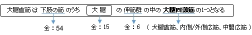
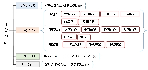
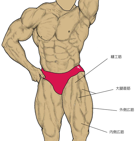
大腿直筋( だいたいちょくきん、英:rectus femoris )
・ 概 要 |
・ 作 用 |
・ イラスト掲載サイト |
|
・ イラスト |
・ 神経 / 脈管 |
||
・ 起始 / 停止 |
・ Wikipedia |
![]()
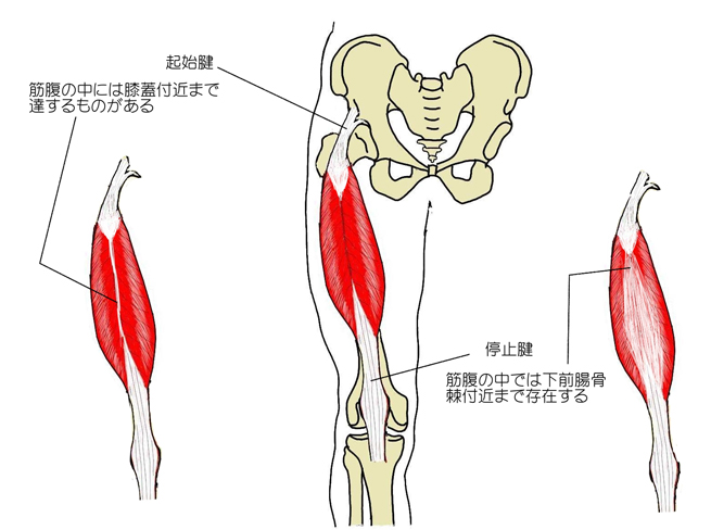
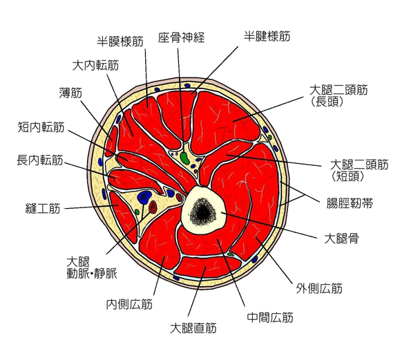
・全体的には紡錘状を呈するが、表面からは羽状の筋に見える。
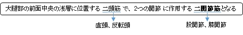
・大腿四頭筋の中で唯一の2関節筋となる。
・腱は起始部も停止部もそれぞれ表面に見える部分だけでなく筋腹の中まで存在し、その一部は起始部は停止部付近まで、停止部は起始部付近まで伸びているものがある。 ⇒ イラスト解説
「骨格筋の形と触察法」のP213から214にかけて以下のような記述がある。
「一般に中間広筋、内側広筋および外側広筋の共通の停止腱の深層に存在し、その内面に終わる筋腹を中間広筋といい、停止腱の浅層に存在し、その外面に終わる筋腹を内側広筋または外側広筋という。しかし、一般的に外側広筋の斜頭といわれる部分の筋線維の一部は、停止腱の内面に終わる。また、停止腱は2~3層に分かれる場合も多く、中間広筋は内側広筋や外側広筋と区別しにくい。」
![]()
 |
 |
 |
 |
||
 |
|
||||
![]()
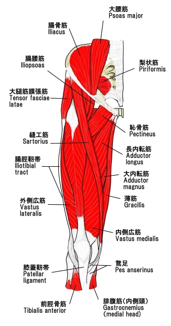
【 起 始 】 : 下前腸骨棘(腸骨)、寛骨臼(腸骨)の上縁
【 停 止 】 : 膝蓋骨の骨底、脛骨粗面(脛骨)(一部が膝蓋靭帯を介して)
※資料によっては停止部を膝蓋骨で留めているものも多い。
![]()
「 大腿の挙上。起立の姿勢では骨盤を屈曲」 ( 日本人体解剖学 )
※二関節筋で膝関節にも作用するので 「膝関節を伸展させる」 という働きも有すると思われる。
![]()
・ 神 経 : 大腿神経(L2,L3,L4)
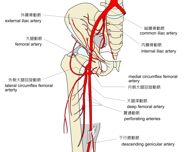
・ 脈 管 : 外側大腿回旋動脈
 |
 |
![]()
The rectus femoris muscle is one of the four quadriceps muscles of the human body. The others are the vastus medialis, the vastus intermedius (deep to the rectus femoris), and the vastus lateralis. All four parts of the quadriceps muscle attach to the patella (knee cap) by the quadriceps tendon.
The rectus femoris is situated in the middle of the front of the thigh; it is fusiform in shape, and its superficial fibers are arranged in a bipenniform manner, the deep fibers running straight (Latin: rectus) down to the deep aponeurosis. Its functions are to flex the thigh at the hip joint and to extend the leg at the knee joint.
【Structure】
It arises by two tendons: one, the anterior or straight, from the anterior inferior iliac spine; the other, the posterior or reflected, from a groove above the rim of the acetabulum.
The two unite at an acute angle and spread into an aponeurosis that is prolonged downward on the anterior surface of the muscle, and from this the muscular fibers arise.
The muscle ends in a broad and thick aponeurosis that occupies the lower two-thirds of its posterior surface, and, gradually becoming narrowed into a flattened tendon, is inserted into the base of the patella.
【 語 句 】
・quadriceps muscles:(大腿)四頭筋 ・vastus medialis:内側広筋 ・vastus intermedius:中間広筋 ・vastus lateralis:外側広筋 ・patella:膝蓋骨 ・quadriceps tendon:四頭筋腱? ・fusiform:紡錘状の ・bipenniform:双翼状の ・aponeurosis:腱膜 ・anterior inferior iliac spine:下前腸骨棘 ・groove:溝 ・acetabulum:寛骨臼 ・acute angle:鋭角
【Nerve supply】
The neurons for voluntary thigh contraction originate near the summit of the medial side of the precentral gyrus (the primary motor area of the brain). These neurons send a nerve signal that is carried by the corticospinal tract down the brainstem and spinal cord. The signal starts with the upper motor neurons carrying the signal from the precentral gyrus down through the internal capsule, through the cerebral peduncle, and into the medulla. In the medullary pyramid, the corticospinal tract decussates and becomes the lateral corticospinal tract. The nerve signal will continue down the lateral corticospinal tract until it reaches spinal nerve L4. At this point, the nerve signal will synapse from the upper motor neurons to the lower motor neurons. The signal will travel through the anterior root of L4 and into the anterior rami of the L4 nerve, leaving the spinal cord through the lumbar plexus. The posterior division of the L4 root is the Femoral nerve. The femoral nerve innervates the quadriceps femoris, a fourth of which is the rectus femoris. When the rectus femoris receives the signal that has traveled all the way from the medial side of the precentral gyrus, it contracts, extending the knee and flexing the thigh at the hip.
【Function】
The rectus femoris, sartorius, and iliopsoas are the flexors of the thigh at the hip. The rectus femoris is a weaker hip flexor when the knee is extended because it is already shortened and thus suffers from active insufficiency; the action will recruit more iliacus, psoas major, tensor fasciae latae, and the remaining hip flexors than it will the rectus femoris.
Similarly, the rectus femoris is not dominant in knee extension when the hip is flexed since it is already shortened and thus suffers from active insufficiency. In essence: the action of extending the knee from a seated position is primarily driven by the vastus lateralis, vastus medialis, and vastus intermedius, and less by the rectus femoris.
In the other extreme, the muscle's ability to flex the hip and extend the knee can be compromised in a position of full hip extension and knee flexion, due to passive insufficiency.
The rectus femoris is a direct antagonist to the hamstrings, at the hip and at the knee.
【 語 句 】
・voluntary:随意の ・contraction:収縮 ・precentral gyrus:中心前回 ・primary motor area:一次運動野 corticospinal tract・:皮質脊髄路 ・brainstem:脳幹 ・internal capsule:内包? ・cerebral peduncle:大脳脚 ・medulla:延髄 ・decussate:X字形の ・lateral corticospinal tract:外側皮質脊髄路 ・anterior root:前根 ・anterior rami :前枝 ・lumbar plexus:腰神経叢 ・contract:収縮する ・sartorius:縫工筋 ・iliopsoas:腸腰筋 ・insufficiency:不十分、機能不全 ・iliacus:腸骨筋 ・psoas major:大腰筋 ・tensor fasciae latae:大腿筋膜張筋 ・dominant:最も有力な ・in essence:本質的に ・antagonist:亀甲均 ・hamstrings:ハムストリングス
![]()
![]()