

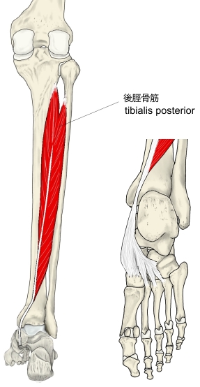
後脛骨筋 ( こうけいこつきん、英:tibialis posterior muscle )
・ 概 要 |
・ 作 用 |
・ イラスト掲載サイ |
|
・ イラスト |
・ 神経 / 脈管 |
||
・ 起始 / 停止 |
・ Wikipedia |
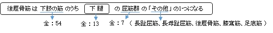
・「足底のアーチを作る筋肉の一つ」とも言われている。
・「骨格筋の形と触察法」には以下のような説明が見受けられる。
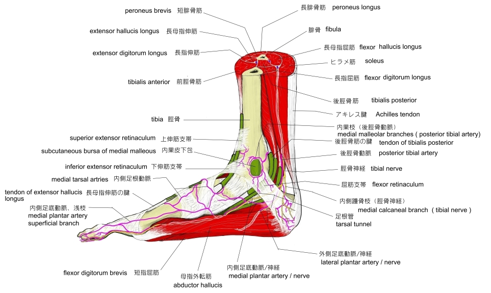
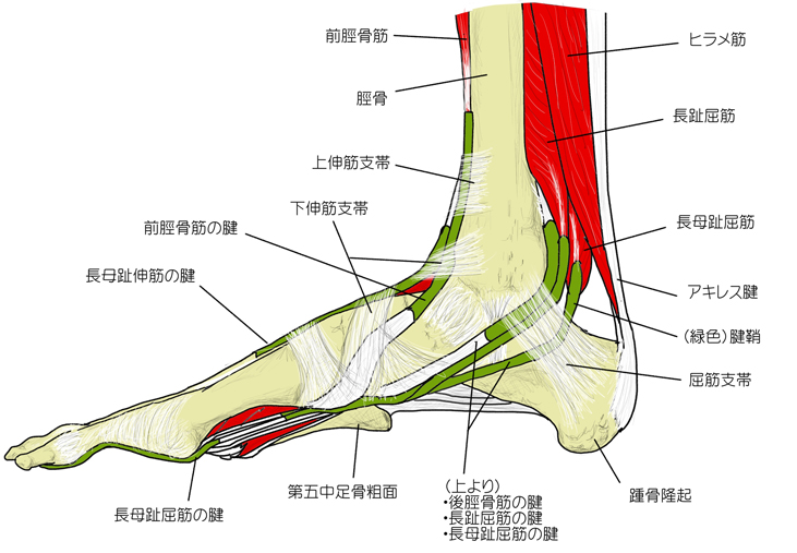
「後脛骨筋の腱は、脛骨の内果の頭方で長趾屈筋の腱の深層を内側方に横切る。」 ⇒ イラスト
筋連結 : 長趾屈筋、長母趾屈筋、ヒラメ筋、前脛骨筋、長母趾伸筋、膝窩筋
以下は「船戸和也のHP」の解説文となる。
「後脛骨筋は骨間膜の広い領域から起こる。狭い辺縁部は腓骨と脛骨の近位部に起こり、浅い線維束は浅深の屈筋間にある結合組織から起こる。後脛骨筋の腱は内果上方で長趾屈筋の腱の下を横切り(下腿腱交叉)、その主束は舟状骨粗面につき、その外側束は(しばしば)すべての遠位足根骨と第2~4中足骨底の足底面に付く。内果溝で後脛骨筋の腱は腱鞘に包まれ、内果下方では屈筋支帯におおわれている。深屈筋群へ脛骨神経の筋枝の中で後脛骨筋への枝はほかの枝よりもずっと近位、ヒラメ筋腱弓のレベルで出る。母趾とそのほかの趾への長屈筋群に対する近位の枝は下腿中位1/3へうつるレベルで脛骨神経から分かれる。脛骨神経からの筋枝は普通下腿遠位1/2からも分かれる。後脛骨筋は脛骨神経の支配を受ける。この筋の収縮により距腿関節における足の底屈、距骨下関節および横足根関節による足の内反が得られる。この筋は内層の縦足弓を維持するうえにも重要である。この筋の収縮が足底で数個の骨を互いに引き寄せるうえに役立つことにも注意すべきである。)」
 |
 |
 |
|||
 |
|||||
【 停 止 】: 舟状骨粗面、内側/中間/外側楔状骨、立方骨、第2~第4中足骨(底側面)
※「プロメテウス解剖学アトラス/解剖学総論・運動器系」では立方骨は含めていない。
 |
「足を足底側に屈曲し、同時に内反する。 」 ( 日本人体解剖学 )
・ 神 経 : 脛骨神経(L5,S1,S2)
The tibialis posterior muscle is the most central of all the leg muscles, and is located in the deep posterior compartment of the leg. It is the key stabilizing muscle of the lower leg.
【Structure】
The tibialis posterior muscle originates on the inner posterior border of the fibula laterally. It is also attached to the interosseous membrane medially, which attaches to the tibia and fibula.
The tendon of the tibialis posterior muscle (sometimes called the posterior tibial tendon) descends posterior to the medial malleolus. It terminates by dividing into plantar, main, and recurrent components. The main portion inserts into the tuberosity of the navicular bone.The smaller portion inserts into the plantar surface of the medial cuneiform. The plantar portion inserts into the bases of the second, third and fourth metatarsals, the intermediate and lateral cuneiforms and the cuboid. The recurrent portion inserts into the sustentaculum tali of the calcaneus.
Blood is supplied to the muscle by the posterior tibial artery.
【Nerve supply】
The tibialis posterior muscle is suppled by the tibial nerve.
【Function】
The tibialis posterior muscle is a key muscle for stabilization of the lower leg. It also contracts to produce inversion of the foot, and assists in the plantarflexion of the foot at the ankle. The tibialis posterior has a major role in supporting the medial arch of the foot. Dysfunction of the tibialis posterior, including rupture of the tibialis posterior tendon, can lead to flat feet in adults, as well as a valgus deformity due to unopposed eversion when inversion is lost.
【 語 句 】
・fibula:腓骨 ・interosseous membrane:骨間膜 ・tibia:脛骨 ・medial malleolus:内果 ・tuberosity:結節 ・navicular bone:舟状骨 ・medial cuneiform:内側楔状骨 ・metatarsal:中足骨 ・cuboid:立方骨 ・sustentaculum tali:載距突起 ・calcaneus:踵骨 ・posterior tibial artery:後脛骨動脈 ・tibial nerve:脛骨神経 ・inversion:転回、反転 ・dysfunction:機能障害 ・rupture:断裂 ・valgus deformity:外反変形 ・unopposed:対立しない ・eversion:外転、外反
![]()