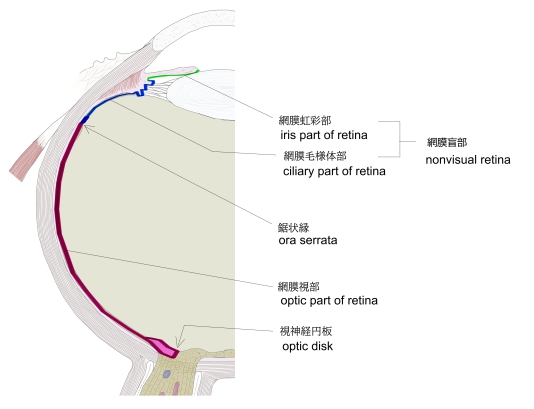
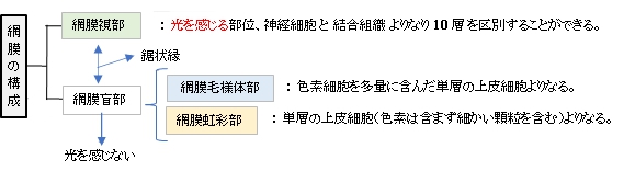
・「網膜視部」に対して前部(毛様体部・虹彩部)の光を感じない部分を網膜盲部と呼ぶ。
・「生体では硝子様透明、平滑であるが、死後は不透明となりヒダを生じる。」(日本人体解剖学)
・「生体では硝子様透明、平滑であるが、死後は不透明となりヒダを生じる」(日本人体解剖学)


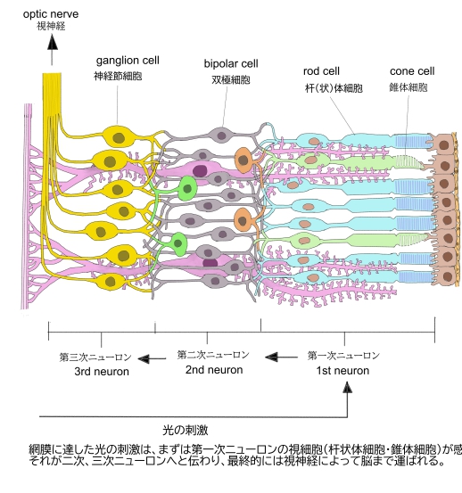
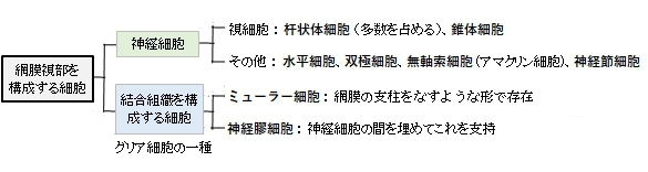
網膜視部に見られる細胞は、大きく情報の伝達を行う神経細胞と結合組織を構成する細胞の2つに分けることができる。

「日本人体解剖学」では網膜視部を外方から内方に向かって以下の10層構造としている。
1 |
杆状体・
錐状体叢層 |
layer of rods and cones
視細胞(杆(状)体細胞および錐体細胞)の外節すなわち杆状体および錐状体の突起部からできる。 |
2 |
外境界膜 |
outer limiting membrane
内境界膜と同じく、ミュラーの支持細胞の終足からなる層 |
3 |
外顆粒層 |
outer granular layer
杆(状)体細胞および錐体細胞からできている。両視細胞は核をもち、2本の突起を持っている。その1突起は杆状体繊維または錐状体線維となって外網状層にのび、そこで内顆粒層の双極神経細胞の突起と連絡する。他の突起は反対の方向に進んで杆状体および錐状体と呼ばれ、杆状体・錐状体層にのびる。 |
4 |
ヘンレ線維層 |
Henle’s outer fiber layer
斜めに走る杆状体および錐状体の線維からできている。
なお、ヘンレ線維層は外網状層の一部として、独立した層として扱わない場合もある。 |
5 |
外網状層 |
outer plexiform layer
杆(状)体細胞および錐体細胞の突起、双極神経細胞の樹状突起が主なものである。そのほか、水平細胞、無軸索細胞(アマクリン細胞)、ミュラーの支持細胞の線維を含む。 |
6 |
内顆粒層 |
inner granular layer
双極神経細胞の数層からなり、この神経細胞の突起は内・外網状層に分枝し終わっている。そのほか、無軸索細胞(アマクリン細胞)、ミュラーの支持細胞を含む。
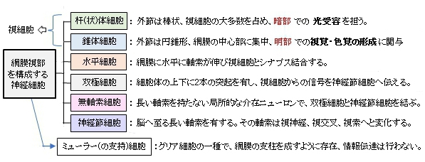
・双極神経細胞(bipolar cell)
細胞は1つの突起を外方すなわち外網状層に送り、その終末によって視細胞とシナプスし、またもう1つの突起を内方すなわち内網状層に送って多極神経細胞とシナプスする。
・無軸索細胞 (アマクリン細胞 amacrine cell)
顆粒層の最内層にある短い突起をもつ細胞で、双極神経細胞とシナプスする。
・水平細胞 horizontal cell
内顆粒層の外帯には水平細胞があって、突起によって視細胞とシナプスする。水平細胞は視細胞間を連合するものと考えられている。
ミュラーの支持細胞 (Muller’s sustentacular cell)
特別な形をした神経膠細胞である。細胞は長くのび、網膜の外境界膜から内境界膜を放線状に貫いている。 |
7 |
内網状層 |
inner plexiform layer
内顆粒層の双極神経細胞の軸索突起、無軸索細胞やミュラーの支持細胞の突起、および視神経細胞の樹状突起などからできている。 |
8 |
神経細胞層 |
layer of ganglion cells
多極神経細胞(視神経細胞)の単層からでき、これからの軸索は視神経線維層の主部をなし、また樹状突起は内網状層の構成に預かっている。 |
9 |
神経線維層 |
layer of optic nerve fibers
多極神経細胞からくる軸索からでき、視神経を構成し脳に向かう。 |
10 |
内境界膜 |
inner limiting membrane
ミュラーの支持細胞の終足からなる層。
網膜内のニューロン:網膜視部には、視細胞、双極神経細胞、多極神経細胞(視神経細胞)の3つのニューロンを区別する。 |

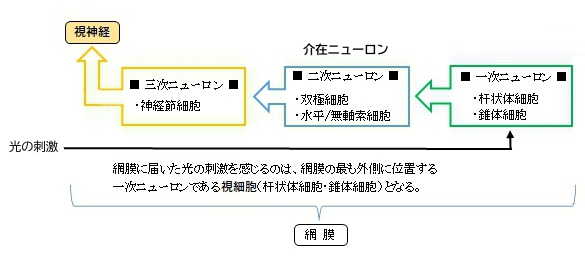
網膜視部に届いた光の刺激は、まず網膜視部の一番外側に位置する一次ニューロンである杆状体細胞および錐体細胞
で感じ取られ、そこから二次ニューロン、そして三次ニューロンへと伝達していく。

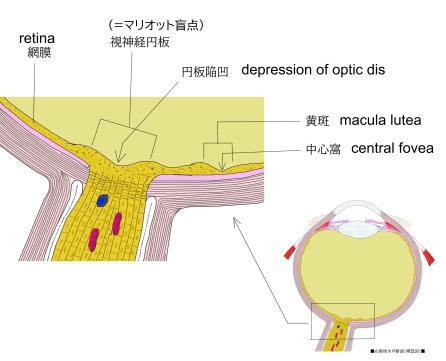
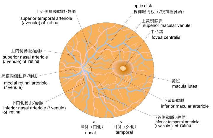
「 日本人体解剖学 」では以下の3つを網膜の特異な部位としている。
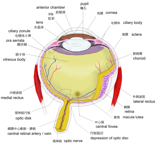
1. 視神経円板 : 眼球を内腔から見たときに視神経が進入する楕円形の部位
2. 黄斑 : 視神経円板の外側に位置する視覚が最も鋭敏な部分
3. 鋸状縁 : 網膜視部と網膜盲部の境界となる非常に薄い部分
|
|
|
|
眼球・視神経進入部 |
右眼球(横断面) |
左眼球・眼底 |
視神経円板・黄斑 |

あ行 |
え |
|
お |
|
か行 |
き |
|
さ行 |
し
|
|
た行 |
ち |
|
な行 |
に |
|
ま行 |
み |
|

以下は「Wikipedia」の「retina」の解説文の一部となる。
□Inverted versus non-inverted retina□
The vertebrate retina is inverted in the sense that the light-sensing cells are in the back of the retina, so that light has to pass through layers of neurons and capillaries before it reaches the photosensitive sections of the rods and cones. The ganglion cells, whose axons form the optic nerve, are at the front of the retina; therefore, the optic nerve must cross through the retina en route to the brain. No photoreceptors are in this region, giving rise to the blind spot. In contrast, in the cephalopod retina, the photoreceptors are in front, with processing neurons and capillaries behind them. Because of this, cephalopods do not have a blind spot.
【 語 句 】
・inverted:逆になった? ・capillary:毛細血管 ・photosensitive:感光性の ・en route to~:~への途中 ・blind spot:盲点 ・cephalapod:(タコ・イカなどの)頭足綱の動物
Although the overlying neural tissue is partly transparent, and the accompanying glial cells have been shown to act as fibre-optic channels to transport photons directly to the photoreceptors, light scattering does occur. Some vertebrates, including humans, have an area of the central retina adapted for high-acuity vision. This area, termed the fovea centralis, is avascular (does not have blood vessels), and has minimal neural tissue in front of the photoreceptors, thereby minimizing light scattering.
【 語 句 】
・transparent: ・glial cell: ・fibre-optic:光ファイバーの ・photon: ・light scattering:光散乱 ・acuity:鋭敏さ ・fovea centralis:中心窩 ・avascular:無血管の
The cephalopods have a non-inverted retina, which is comparable in resolving power to the eyes of many vertebrates. Squid eyes do not have an analog of the vertebrate retinal pigment epithelium (RPE). Although their photoreceptors contain a protein, retinochrome, that recycles retinal and replicates one of the functions of the vertebrate RPE, cephalopod photoreceptors are likely not maintained as well as in vertebrates, and that as a result, the useful lifetime of photoreceptors in invertebrates is much shorter than in vertebrates. Having easily replaced stalk eyes (some lobsters) or retinae (some spiders, such as Deinopis) rarely occurs.
The cephalopod retina does not originate as an outgrowth of the brain, as the vertebrate one does. This difference suggests that vertebrate and cephalopod eyes are not homologous, but have evolved separately.
【 語 句 】
・resolving power:解像力 ・analog:相似器官 ・retinal pigmental epithelium:網膜色素上皮 ・retinochrome:レチノクロム(感光性色素蛋白)) ・replicate:模写する ・stalk:細長い支え ・homologous:相同の ・evolve:進化する
From an evolutionary perspective, a more complex structure such as the inverted retina can generally come about as a consequence of two alternate processes - an advantageous "good" compromise between competing functional limitations, or as a historical maladaptive relic of the convoluted path of organ evolution and transformation. Vision is an important adaptation in higher vertebrates.
A third view of the "inverted" vertebrate eye is that it combines two benefits - the maintenance of the photoreceptors mentioned above, and the reduction in light intensity necessary to avoid blinding the photoreceptors, which are based on the extremely sensitive eyes of the ancestors of modern hagfish (fish that live in very deep, dark water).
【 語 句 】
・perspective:考え方 ・come about:起こる ・consequence:結果、結論 ・alternate:交互の ・compromise:妥協 ・maladaptive:不適応の ・convoluted:回旋状の、入り組んだ ・adaptation:適応 ・hagfish:メクラウナギ
■ 写真やイラストを掲載しているサイト ■
・ イラストや写真を掲載しているサイト-Ⅰ
・ イラストや写真を掲載しているサイト-Ⅱ
・ イラストや写真を掲載しているサイト-Ⅲ
・ イラストや写真を掲載しているサイト-Ⅳ
・ イラストや写真を掲載しているサイト-Ⅴ