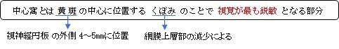
・「中心窩は血管のない黄斑の中心にあるくぼみ。」(船戸和也のHP)
・「中心窩は視力が最も良く、ここでいる視力を中心視力という。」(船戸和也のHP)
・「中心窩の位置はほぼ眼球の後極に当たっている.」(Rauber-Kopsh解剖学)
・「中心窩の中心部をfoveola(小窩)といい錐体細胞(cone cell)のみで構成される。」(Wikipedia)
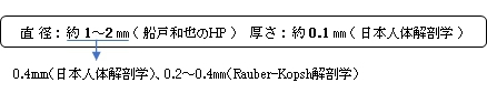
※直径は資料によってかなり違いが見られる。おそらく「anatomical」と「clinical」によるものだと思われる。
|
|
|
|
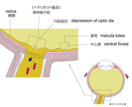
眼球・視神経進入部 |
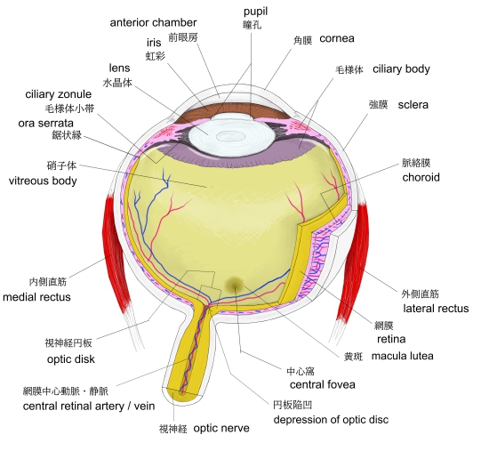
右眼球(横断面) |
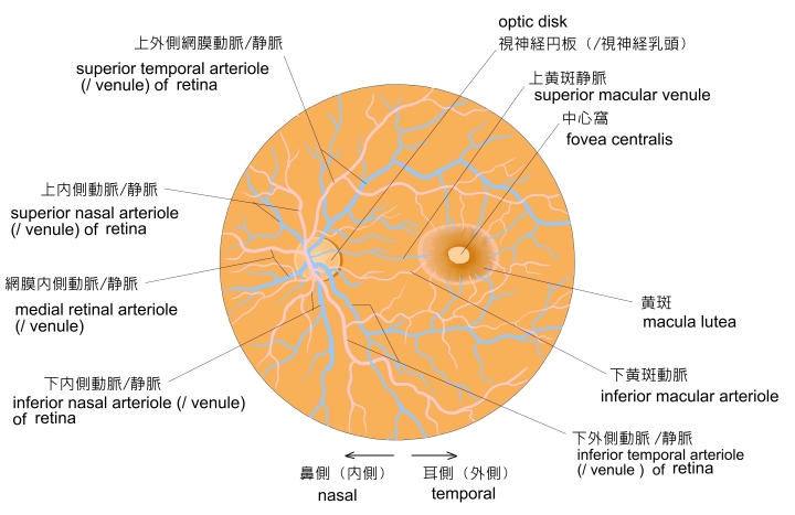
左眼球・眼底 |
視神経円板・黄斑 |
以下は「Rauber-Kopsh解剖学」の解説文の一部となる。
「新鮮な眼では,したがってまた生体の眼底鏡像では,黄斑および中心窩のところが黄色ではなくて,赤褐色
ないし褐色にみえ,中心窩のところは薄くて透明なので,その下にある層がすき通ってかすかにみえる.」
また、以下は「Wikipedia」の解説文となる。
The fovea centralis is a small, central pit composed of closely packed cones in the eye. It is located in the center of the macula lutea of the retina.
The fovea is responsible for sharp central vision (also called foveal vision), which is necessary in humans for activities for which visual detail is of primary importance, such as reading and driving. The fovea is surrounded by the parafovea belt and the perifovea outer region.
The parafovea is the intermediate belt, where the ganglion cell layer is composed of more than five layers of cells, as well as the highest density of cones; the perifovea is the outermost region where the ganglion cell layer contains two to four layers of cells, and is where visual acuity is below the optimum. The perifovea contains an even more diminished density of cones, having 12 per 100 micrometres versus 50 per 100 micrometres in the most central fovea. That, in turn, is surrounded by a larger peripheral area, which delivers highly compressed information of low resolution following the pattern of compression in foveated imaging.
Approximately half the nerve fibers in the optic nerve carry information from the fovea, while the remaining half carry information from the rest of the retina. The parafovea extends to a radius of 1.25 mm from the central fovea, and the perifovea is found at a 2.75 mm radius from the fovea centralis.
【 語 句 】
・cone:錐体細胞 ・macula lutea:黄斑 ・retina:網膜 ・parafovea:傍中居窩 ・perifovea:周中心窩 ・ganglion cell:神経節細胞 ・optimum:最適条件 ・diminished:減少して ・peripheral:周囲の ・compressed:圧縮した ・low resolution:低分解能 ・foveated:中心窩で捉える ・radius:半径
【Structure】
The fovea is a depression in the inner retinal surface, about 1.5 mm wide, the photoreceptor layer of which is entirely cones and which is specialized for maximum visual acuity. Within the fovea is a region of 0.5mm diameter called the foveal avascular zone (an area without any blood vessels). This allows the light to be sensed without any dispersion or loss. This anatomy is responsible for the depression in the center of the fovea. The foveal pit is surrounded by the foveal rim that contains the neurons displaced from the pit. This is the thickest part of the retina.
The fovea is located in a small avascular zone and receives most of its oxygen from the vessels in the choroid, which is across the retinal pigment epithelium and Bruch's membrane. The high spatial density of cones along with the absence of blood vessels at the fovea accounts for the high visual acuity capability at the fovea.
The center of the fovea is the foveola – about 0.35 mm in diameter – or central pit where only cone photoreceptors are present and there are virtually no rods.The central fovea consists of very compact cones, thinner and more rod-like in appearance than cones elsewhere. These cones are very densely packed (in a hexagonal pattern). Starting at the outskirts of the fovea, however, rods gradually appear, and the absolute density of cone receptors progressively decreases.
In 2018 the anatomy of the foveola was reinvestigated, and it was discovered that outer segments from the central foveolar cones of monkeys are not straight and twice as long as those from the parafovea.
【Size】
The size of the fovea is relatively small with regard to the rest of the retina. However, it is the only area in the retina where 20/20 vision is attainable, and is the area where fine detail and colour can be distinguished.
【 語 句 】
・photoreceptor:光受容器、視細胞 ・acuity:鋭敏さ ・foveal avascular zone:中心窩無血管領域 ・dispersion:分散・choroid:脈絡膜 ・retinal pigment epithelium:網膜色素上皮 ・Bruch's membrane:ブルッフ膜 ・spatial:空間の ・foveola:小窩 ・rod:杆状体細胞 ・hexagonal:六角形の ・progressively:次第に・reinvestigate:再調査する ・attainable:到達できる
■ 写真やイラストを掲載しているサイト ■
・ イラストや写真を掲載しているサイト-Ⅰ
・ イラストや写真を掲載しているサイト-Ⅱ
・ イラストや写真を掲載しているサイト-Ⅲ
・ イラストや写真を掲載しているサイト-Ⅳ
・ イラストや写真を掲載しているサイト-Ⅴ