・「神経細胞層の重層からできる。内顆粒層は著しく発達し、杆状体細胞を欠く」(日本人体解剖学)
・Wikipediaでは黄斑を以下のように6つの部位に細分化している。※日本語訳は正確性に欠ける可能性がある。
1.umbo(殻頂?) 2.foveola(小窩) 3.foveal avascular zone(中心窩無血管領域)
4.fovea(中心窩) 5.parafovea(傍中居窩) 6.perifovea(周中心窩)
注意)Wikipediaでは黄斑を「anatomical(解剖学の)」と「clinical(臨床の)」の2通りの区別をしている。
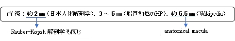
そして、その直径もclinicalでは「1.5㎜」としている。よって、上の直径約2㎜というのは、Wikipediaでいう
clinicalのサイズをいうものなのかもしれない。

|
|
|
|
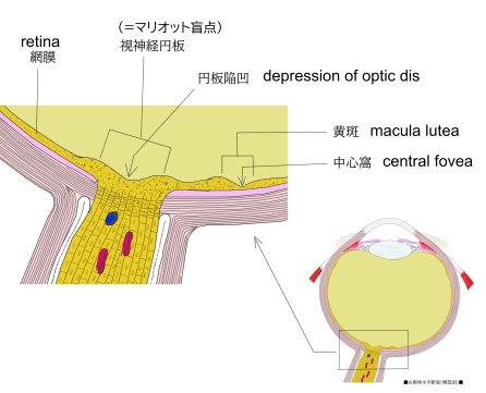
眼球・視神経進入部 |
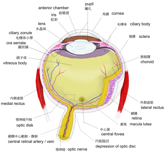
右眼球(横断面) |
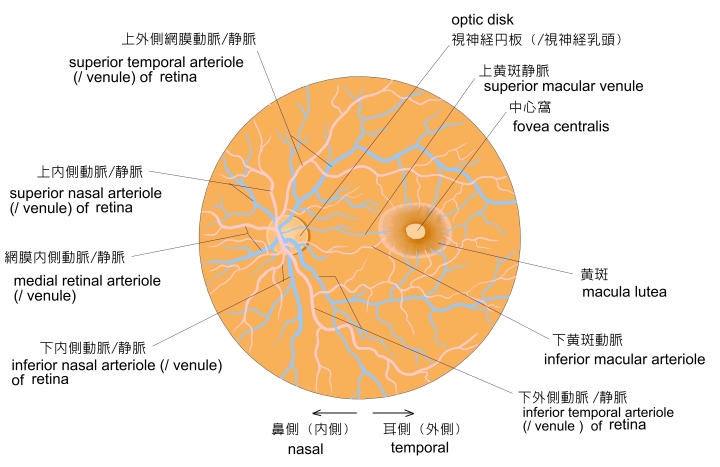
左眼球・眼底 |
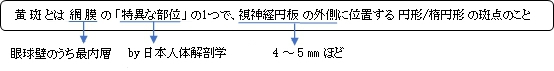
視神経円板・黄斑 |
以下は「Wikipedia」の解説文となる。
The macula or macula lutea is an oval-shaped pigmented area in the center of the retina of the human eye and in other animals. The macula in humans has a diameter of around 5.5 mm (0.22 in) and is subdivided into the umbo, foveola, foveal avascular zone, fovea, parafovea, and perifovea areas.
The anatomical macula at a size of 5.5 mm (0.22 in) is much larger than the clinical macula which, at a size of 1.5 mm (0.059 in), corresponds to the anatomical fovea.
The macula is responsible for the central, high-resolution, color vision that is possible in good light; and this kind of vision is impaired if the macula is damaged, for example in macular degeneration. The clinical macula is seen when viewed from the pupil, as in ophthalmoscopy or retinal photography.
The term macula lutea comes from Latin macula, "spot", and lutea, "yellow".
【 語 句 】
・pigmented:色素沈着 ・retina:網膜 ・subdivide:細分する ・umbo:殻頂? ・foveola:小窩 ・foveal avaschular zone:中心窩無血管領域 ・fovea:中心窩 ・parafovea:傍中居窩 ・perifovea
周中心窩: ・anatomical:解剖学の ・clinical:臨床の ・impair:害する ・macular degeneration:黄斑変性症 ・pupil:瞳孔 ・ophthalmoscopy:眼底検査 ・retinal photography:網膜写真撮影
【
Structure】
Photograph of the retina of the human eye, with overlay
diagrams showing the positions and sizes of the macula, fovea, and optic disc
Schematic diagram of the macula lutea of the retina, showing perifovea, parafovea, fovea, and clinical macula
The macula is an oval-shaped pigmented area in the center of the
retina of the
human eye and other animal
eyes. Its center is shifted slightly away from the
optical axis (laterally, by 5°=1.5 mm). The macula in humans has a diameter of around 5.5 mm (0.22 in) and is subdivided into the
umbo,
foveola,
foveal avascular zone,
fovea,
parafovea, and
perifovea areas. An even smaller central region of highest receptor density (40–80 μm) is sometimes referred to as the
foveal bouquet. The anatomical macula at 5.5 mm (0.22 in) is much larger than the clinical macula which, at 1.5 mm (0.059 in),
corresponds to the anatomical fovea.
The clinical macula is seen when viewed from the pupil, as in
ophthalmoscopy or
retinal photography. The anatomical macula is
defined histologically in terms of having two or more layers of
ganglion cells. The umbo is the center of the foveola which in turn is located at the center of the fovea.
The fovea is located near the center of the macula. It is a small pit that contains the largest concentration of
cone cells. The retina's receptor layer contains two types of
photosensitive cells, the
rod cells and the cone cells.
【 語 句 】
・diagram:図形、図解、図表 ・Schematic diagram:模式図 ・optical axis:光軸 ・correspond to ~:~に対応して ・define:定義する ・ganglion cell:神経節細胞 ・cone cell:錐体細胞 ・photosensitive cells:感光細胞? ・rod cell:桿状(体)細胞
【Color】
Because the macula is yellow in color it absorbs excess blue and ultraviolet light that enter the eye and acts as a natural sunblock (analogous to sunglasses) for this area of the retina. The yellow color comes from its content of lutein and zeaxanthin, which are yellow xanthophyll carotenoids, derived from the diet. Zeaxanthin predominates at the macula, while lutein predominates elsewhere in the retina. There is some evidence that these carotenoids protect the pigmented region from some types of macular degeneration. A formulation of 10 mg lutein and 2 mg zeaxanthin has been shown to reduce the risk of age-related macular degeneration progressing to advanced stages, although these carotenoids have not been shown to prevent the disease.
After death or enucleation (removal of the eye), the macula appears yellow, a color that is not visible in the living eye except when viewed with light from which red has been filtered.
【Regions】
【 語 句 】
・exess:超過の、余分の ・ultraviolet light:紫外線 ・analogous:類似して ・lutein:ルテイン ・zeaxanthin:ゼアキサンチン ・xanthophyll:キサントフィル ・carotenoid:カロチノイド ・predominate:優位を占める ・degeneration:変性、変質 ・enucleation:眼球除去
【Function】
Structures in the macula are specialized for high-acuity vision. Within the macula are the fovea and foveola that both contain a high density of cones, which are nerve cells that are photoreceptors with high acuity.
In detail, the normal human eye contains three different types of cones, with different ranges of spectral sensitivity. The brain combines the signals from neighboring cones to distinguish different colors. There is only one type of rod, but the rods are more sensitive than the cones, so in dim light, they are the dominant photoreceptors active, and without information provided by the separate spectral sensitivity of the cones it is impossible to discriminate colors. In the fovea centralis, cones predominate and are present at high density. The macula is thus responsible for the central, high-resolution, color vision that is possible in good light; and this kind of vision is impaired if the macula is damaged, for example in macular degeneration.
【 語 句 】
・acuity:鋭敏さ ・photoreceptor:光受容細胞 ・spectral:スペクトルの ・sensitivity:感光度 ・distinguish:識別する ・dim:ほの暗い ・discriminate:区別する ・impair:減じる、害する ・macular degeneration:黄斑変性症
■ 写真やイラストを掲載しているサイト ■
・ イラストや写真を掲載しているサイト-Ⅰ
・ イラストや写真を掲載しているサイト-Ⅱ
・ イラストや写真を掲載しているサイト-Ⅲ
・ イラストや写真を掲載しているサイト-Ⅳ
・ イラストや写真を掲載しているサイト-Ⅴ