

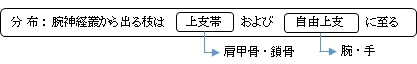
「 第1肋骨と前斜角筋、中斜角筋の間(斜角筋隙)を通り、鎖骨の後上方から下外側方すなわち腋窩付近にわたる脊髄神経叢で、これから出る枝は上肢帯および自由上肢帯に至る。 」(日本人体解剖学 (上巻) ) )
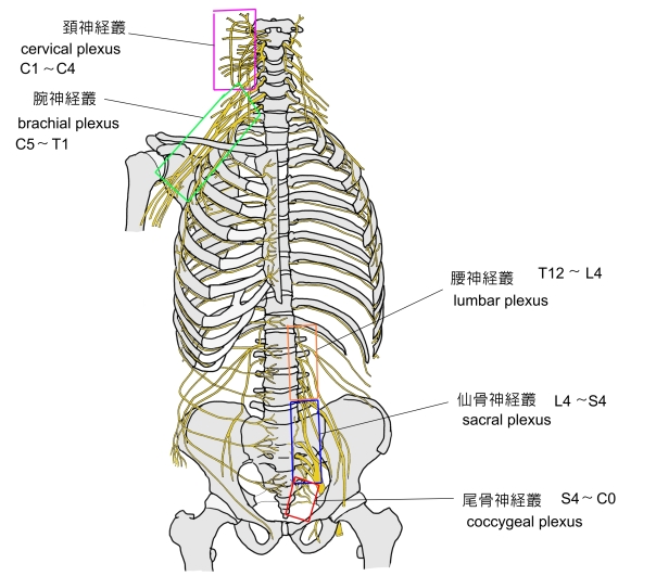
以下は脊髄神経が形成する神経叢を簡単に表した図になる。
※「 日本人体解剖学 (上巻) 」では、陰部神経叢を仙骨神経叢に含めて解説している。

⇒ イラスト解説
「 日本人体解剖学 (上巻) 」では以下のように解説している。
「第1肋骨と前斜角筋、中斜角筋の間(斜角筋隙)を通り、鎖骨の後上方から下外側方すなわち腋窩付近にわたる脊髄神経叢で、これから出る枝は上肢帯および自由上肢帯に至る。 」

■ 神経幹 : nerve trunk ■
「 日本人体解剖学 (上巻) 」には以下のような解説が見られる。
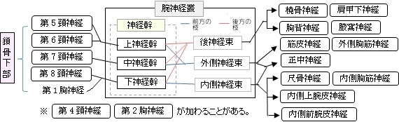
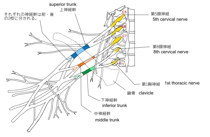
「 腕神経叢の構成様式は、極めて複雑で、多様な変異が見られる。以下、日本人における正常型 (約75%)について述べる。 」

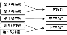
・ 上神経幹 ( superior trunk ): 第5、第6頚神経よりなる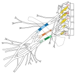
・ 中神経幹 ( middle trunk ) : 第7頚神経よりなる
・ 下神経幹 ( inferior trunk ) : 第8頚神経、第1胸神経よりなる
■ 神経束 : nerve fasciculus (/cord) ■

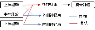
・ 上・中・下神経幹の3つの後枝が合して 後神経束 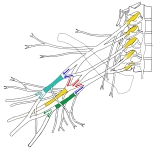 を形成し、そのまま延長して 橈骨神経 となる。 を形成し、そのまま延長して 橈骨神経 となる。
・ 上・中神経幹の前枝が合して 外側神経束 を形成して2つに分岐する。
・ 下神経幹の前枝はそのまま 内側神経束 となり2つに分岐する。
「 日本人体解剖学 (上巻) 」には以下のような解説が見られる。
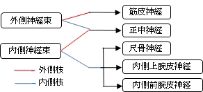
「ついで、外側神経束、内側神経束はおのおの分かれて2枝となり、このようにしてできた4枚のうち、中央の2枝は合っして正中神経を作り、外側神経束の他の1枝すなわち外側枝は筋皮神経、内側神経束の他の1枝すなわち内側枝はさらに3枝に分かれて尺骨神経、内側上腕皮神経、内側前腕皮神経となる。 」


「 日本人体解剖学 (上巻) 」には以下のような解説が見られる。
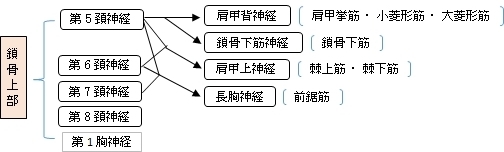
「腕神経叢は、鎖骨によって鎖骨上部と鎖骨下部の2部に分けられる。多くの場合、鎖骨上部では神経幹の形成が、鎖骨下部では内側、外側、後の神経束の形成が行われる。鎖骨上部および鎖骨下部から次の神経が出る。」
■ 鎖骨上部 : supraclavicular branches ■ 
■ 鎖骨下部 : infraclavicular branches ■

以下は「 Wikipedia 」の解説文となる。
「 The brachial plexus is a network of nerves formed by the anterior rami of the lower four cervical nerves and first thoracic nerve (C5, C6, C7, C8, and T1). This plexus extends from the spinal cord, through the cervicoaxillary canal in the neck, over the first rib, and into the armpit. It supplies afferent and efferent nerve fibers to the chest, shoulder, arm and hand.
【 Structure 】
The brachial plexus is divided into five roots, three trunks, six divisions, three anterior and three posterior, three cords, and five branches. There are five "terminal" branches and numerous other "pre-terminal" or "collateral" branches, such as the subscapular nerve, the thoracodorsal nerve, and the long thoracic nerve,[1] that leave the plexus at various points along its length.[2] A common structure used to identify part of the brachial plexus in cadaver dissections is the M or W shape made by the musculocutaneous nerve, lateral cord, median nerve, medial cord, and ulnar nerve.
【 Roots 】
The five roots are the five anterior rami of the spinal nerves, after they have given off their segmental supply to the muscles of the neck. The brachial plexus emerges at five different levels; C5, C6, C7, C8, and T1. C5 and C6 merge to establish the upper trunk, C7 continuously forms the middle trunk, and C8 and T1 merge to establish the lower trunk. Prefixed or postfixed formations in some cases involve C4 or T2, respectively. The dorsal scapular nerve comes from the superior trunk[2] and innervates the rhomboid muscles which retract the scapula. The subclavian nerve originates in both C5 and C6 and innervates the subclavius, a muscle that involves lifting the first ribs during respiration. The long thoracic nerve arises from C5, C6, and C7. This nerve innervates the serratus anterior, which draws the scapula laterally and is the prime mover in all forward-reaching and pushing actions.
【 Trunks 】
These roots merge to form three trunks:
- "superior" or "upper" (C5-C6)
- "middle" (C7)
- "inferior" or "lower" (C8, T1)
- 【 Divisions 】
Each trunk then splits in two, to form six divisions:
- anterior divisions of the upper, middle, and lower trunks
- posterior divisions of the upper, middle, and lower trunks
- when observing the body in an anatomical position, the anterior divisions are superficial to the posterior divisions
- 【 Cords 】
These six divisions regroup to become the three cords or large fiber bundles. The cords are named by their position with respect to the axillary artery.
- The posterior cord is formed from the three posterior divisions of the trunks (C5-C8, T1)
- The lateral cord is formed from the anterior divisions of the upper and middle trunks (C5-C7)
- The medial cord is simply a continuation of the anterior division of the lower trunk (C8, T1)
- 【 Branches 】
The branches are listed below. Most branch from the cords, but a few branch (indicated in italics) directly from earlier structures. The five on the left are considered "terminal branches". These terminal branches are the musculocutaneous nerve, the axillary nerve, the radial nerve, the median nerve, and the ulnar nerve. Due to both emerging from the lateral cord the musculocutaneous nerve and the median nerve are well connected. The musculocutaneous nerve has even been shown to send a branch to the median nerve further connecting them.[1] There have been several variations reported in the branching pattern but these are very rare.[3]」
【 語 句 】
・: ・: ・: ・: ・: ・: ・: ・: ・: ・: ・: ・:
【参考になるサイト】
・イラストや写真を掲載しているサイト-Ⅰ
・イラストや写真を掲載しているサイト-Ⅱ
・イラストや写真を掲載しているサイト-Ⅲ
・イラストや写真を掲載しているサイト-Ⅳ
・イラストや写真を掲載しているサイト-Ⅴ

|


.jpg)