

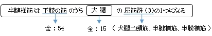
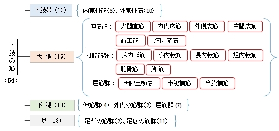
半腱様筋 ( はんけんようきん、英:semitendinosus muscle )
・ 概 要 |
・ 作 用 |
・ イラスト掲載サイト |
|
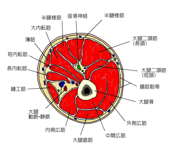
・ イラスト |
・ 神経 / 脈管 |
||
・ 起始 / 停止 |
・ Wikipedia |
![]()
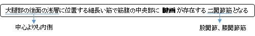
・上部半分の筋腹は厚く、停止付近は主に腱で構成されている(それがこの筋の名称の由来)。
・大腿二頭筋、半膜様筋とともにハムストリングス(膝窩腱)を、薄筋及び縫工筋の腱とともに鵞足を構成する。
筋連結 : 大腿二頭筋(長頭)、半膜様筋、縫工筋、薄筋、大内転筋、 腓腹筋
以下は「船戸和也のHP」の解説文となる。
「半腱様筋は大腿二頭筋長頭の起始近くの坐骨結節から起こり、鵞足を介して脛骨近位端内側面および下腿筋膜に終わる。半腱様筋は半膜様筋によってつくられた溝の中を遠位へ向かう。長い停止腱は大腿部ですでに始まり(ここから“半腱様”の名がつけられた)、鵞足の深層へと放散する。」
![]()
 |
|||||||
 |
|||||||
![]()
【 停 止 】
「脛骨粗面に沿い、薄筋停止部の後下方につき、また下腿筋膜に続く。半腱様筋は脛骨を取り巻いて扇状の腱に終わり、縫工筋および薄筋の腱とともに鵞足をつくる。」( 日本人体解剖学)
「鵞足を介して脛骨近位端内側面および下腿筋膜に終わる。」(船戸和也のHP)
 |
![]()
「下腿を屈曲し、同時に内側方に回す。大腿を固定するときには骨盤を起立させる。 」 ( 日本人体解剖学 )
・「プロメテウス解剖学アトラス」では
「・股関節:伸展、矢状面内での骨盤の安定 ・膝関節:屈曲と内旋」
![]()
・ 神 経 : 脛骨神経(L4,L5,S1,S2)
※その他として内腸骨動脈および膝窩動脈を挙げている資料も見られる。
 |
![]()
Semitendinosus is one of the three muscles that make up the hamstrings muscle group, and it is located at the posterior and medial aspect of the thigh. The semitendinosus is so named due to it having a long tendon of insertion.
The muscle is fusiform and ends a little below the middle of the thigh in a long round tendon which lies along the medial side of the popliteal fossa; it then curves around the medial condyle of the tibia and passes over the medial collateral ligament of the knee-joint, from which it is separated by a bursa, and is inserted into the upper part of the medial surface of the body of the tibia, nearly as far forward as its anterior crest.
The semitendinosus is more superficial than the semimembranosus (with which it shares very close insertion and attachment points). However, because the semimembranosus is wider and flatter than the semitendinosus, it is still possible to palpate the semimembranosus directly.
At its insertion it gives off from its lower border a prolongation to the deep fascia of the leg and lies behind the tendon of the sartorius, and below that of the gracilis, to which it is united. These three tendons form the Pes Anserinus, thus named due to the appearance resembling a webbed “goose’s foot”.
Anatomy
Origin
It arises, by a common tendon origin with the long head of the biceps femoris, from the lower medial facet of the lateral section of the ischial tuberosity. The semitendinosus muscle mainly originates from the medial surface of the tendon of the long head of the biceps femoris, and also originates from the ischial tuberosity with a thin tendon and a muscular part.
【 語 句 】
・hamstrings:ハムストリングス ・fusiform:紡錘状の ・popliteal fossa:膝窩 ・medial condyle:内側顆 ・tibia:脛骨 ・medial collateral ligament:内側側副靭帯 ・bursa:滑液包 ・semimembranosus:半膜様筋 ・palpate:触診する ・prolongation:延長 ・fascia:筋膜 ・sartorius:縫工筋 ・gracilis:薄筋 ・Pes Anserinus:鵞足 ・biceps femoris:大腿二頭筋 ・ischial tuberosity:坐骨結節
Insertion
The semitendinosus tendon inserts at the upper part of the medial surface of the tibia, behind the attachment of sartorius and infero-anterior to the attachment of gracilis.
Nerve Supply
Tibial portion of the sciatic nerve (L5, S1, 2).
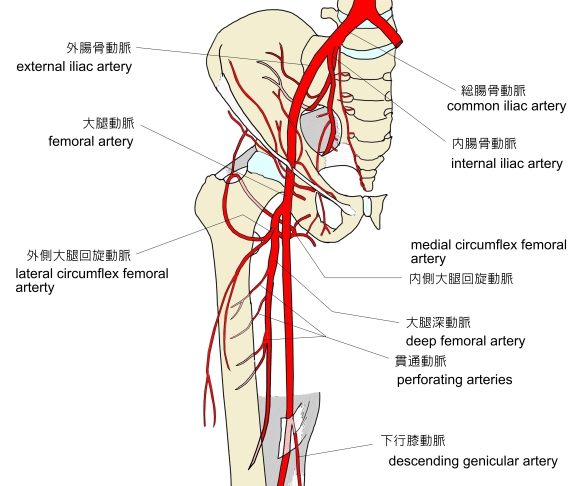
Artery
Branches from the internal iliac, popliteal, and profunda femoris arteries.
Function
1.Extension of the thigh at the hip
- Agonists: gluteus maximus, semimembranosus, biceps femoris (long head), and adductor magnus (posterior part)
- Antagonists: psoas major and iliacus
The semitendinosus is also a weak medial rotator of the hip.
2. Flexion of the leg at the knee
- Agonists: biceps femoris (long head), biceps femoris (short head), and semimembranosus
- Antagonists: vastus lateralis, vastus medialis, vastus intermedius, and rectus femoris
Gracilis, sartorius, popliteus, gastrocnemius, and plantaris assist with flexion of the knee.
3. Internal rotation of the knee when the knee is flexed
- Agonists: popliteus and semimembranosus
- Antagonist: biceps femoris (long head) and biceps femoris (short head)
Sartorius and gracilis assist with internal rotation of knee.
【 語 句 】
・sciatic nerve:坐骨神経 ・internal iliac artery:内腸骨動脈 ・popliteal artery:膝窩動脈 ・profunda femoris artery:大腿深動脈 ・agonist:作動筋 ・gluteus maximus:大殿筋 ・antagonist:拮抗筋 ・psoas major:大腰筋 ・iliacus:腸骨筋 ・vastus lateralis:外側広筋 ・vastus medialis:内側広筋 ・vastus intermedius:中間広筋 ・rectus femoris:大腿直筋 ・gastrocnemius:腓腹筋 ・plantaris:足底筋
![]()
![]()