

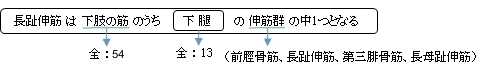
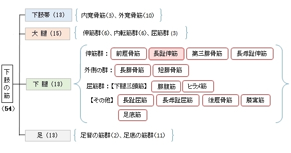
長趾伸筋 ( ちょうししんきん、英:extensor digitorum longus muscle )
・ 概 要 |
・ 作 用 |
・ イラスト掲載サイト |
|
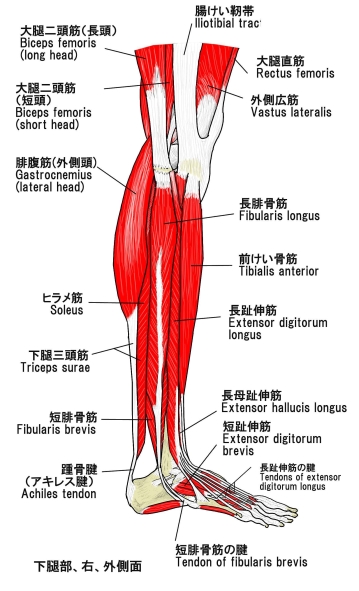
・ イラスト |
・ 神経 / 脈管 |
||
・ 起始 / 停止 |
・ Wikipedia |
![]()
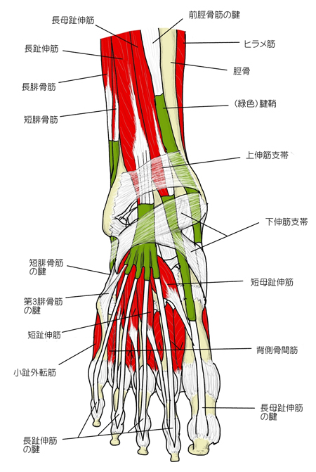
・「 長趾伸筋および第三腓骨筋の停止腱は、足根洞と下伸筋支帯の間にあるワナ靭帯により固定される。」(日本人体解剖学)
筋連結 : 前脛骨筋、長腓骨筋、長母趾伸筋、第三腓骨筋、短腓骨筋、
以下は「船戸和也のHP」の解説文となる。
「長趾伸筋は脛骨外側顆、腓骨前縁および骨間膜の狭い部から起こり、第2~5趾の足背腱膜へ至る。足背腱膜はその基本構造においては手指の手背腱膜と同じである(つまり、各腱の側縁束は末節骨に、中央束は中節骨に終わる)。足背筋膜は趾の部で完全に区分できるとは限らない。骨間膜の腱は通常基節骨にしか停止せず、虫様筋の腱索は第2~5趾の中節骨や末節骨に達するとは限らないので、第2~5趾の各関節を能動的に伸展することはしばしば困難となる。母指末節骨のみは長母趾伸筋の作用によって背屈することが可能である。」
![]()
 |
 |
||||
 |
 |
||||
![]()
【 起 始 】 : 脛骨の外側面の上端、腓骨の前縁、下腿骨間膜、下腿筋膜
【 停 止 】 : 第2~第5趾の指背腱膜
![]()
「 足および第2~第5指を足背側に屈曲し、足の外(脛)側縁を上げる。(外反)足を固定すると、下腿を前方に傾ける。 」 ( 日本人体解剖学 )
![]()
・ 神 経 : 深腓骨神経(L4,L5,S1)
・ 脈 管 : 前脛骨動脈
![]()
The extensor digitorum longus is a pennate muscle, situated at the lateral part of the front of the leg.
【Origin and insertion】
It arises from the lateral condyle of the tibia; from the upper three-quarters of the anterior surface of the body of the fibula; from the upper part of the interosseous membrane; from the deep surface of the fascia; and from the intermuscular septa between it and the tibialis anterior on the medial, and the peroneal muscles on the lateral side. Between it and the tibialis anterior are the upper portions of the anterior tibial vessels and deep peroneal nerve.
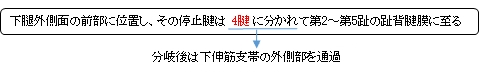
The muscle passes under the superior and inferior extensor retinaculum of foot in company with the fibularis tertius, and divides into four slips, which run forward on the dorsum of the foot, and are inserted into the second and third phalanges of the four lesser toes.
The tendons to the second, third, and fourth toes are each joined, opposite the metatarsophalangeal articulations, on the lateral side by a tendon of the extensor digitorum brevis. The tendons are inserted in the following manner: each receives a fibrous expansion from the interossei and lumbricals, and then spreads out into a broad aponeurosis, which covers the dorsal surface of the first phalanx: this aponeurosis, at the articulation of the first with the second phalanx, divides into three slips—an intermediate, which is inserted into the base of the second phalanx; and two collateral slips, which, after uniting on the dorsal surface of the second phalanx, are continued onward, to be inserted into the base of the third phalanx.
【Variations】
This muscle varies considerably in the modes of origin and the arrangement of its various tendons.
The tendons to the second and fifth toes may be found doubled, or extra slips are given off from one or more tendons to their corresponding metatarsal bones, or to the short extensor, or to one of the interosseous muscles.
A slip to the great toe from the innermost tendon has been found.
【 語 句 】
・pennate:羽毛状の ・lateral condyle:外側顆 ・tibia:脛骨 ・fibula:腓骨 ・interosseous membrane:骨間膜 ・fascia:筋膜 ・intermuscular septa:筋間中隔 ・tibialis anterior:前脛骨筋 ・peroneal muscles:腓骨筋 ・deep peroneal nerve:深腓骨神経 ・inferior extensor retinaculum:下伸筋支帯 ・fibularis tertius:第三腓骨筋 ・phalanges:指骨 ・metatarsophalangeal articulations:中足指節関節 ・extensor digitorum brevis:短趾真菌 ・interossei:interosseus(骨間筋)の複数 ・lumbricals:虫様筋 ・aponeurosis:腱膜 ・metatarsal bones:中足骨 ・interosseous muscles:骨間筋
![]()
![]()