

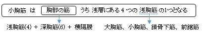
小胸筋 (しょうきょうきん、英:pectoralis minor muscle)
・ 概 要 |
・ 作 用 |
・ イラスト掲載サイト |
|
・ イラスト |
・ 神経 / 脈管 |
||
・ 起始 / 停止 |
・ Wikipedia |
![]()
![]()
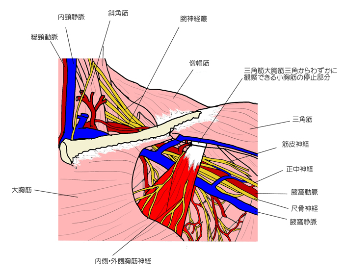
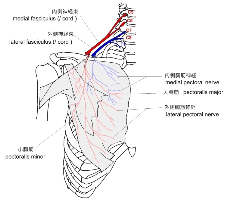
・ 小胸筋の下を 腕神経叢、腋窩動脈、腋窩静脈 が通っている。 ⇒ イラスト
・ 内側胸筋神経が貫通している。
・ 腋窩リンパ節および腋窩動脈は、小胸筋との位置関係によってそれぞれ3部に区別される。
「 船戸和也のHP 」 では以下のように解説している。
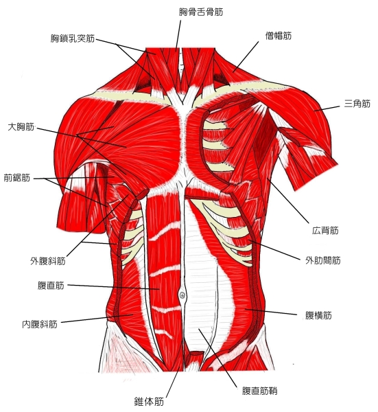
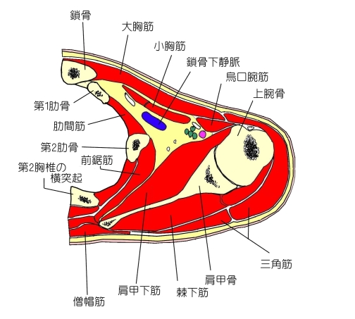
「小胸筋は前胸壁にあり大胸筋で完全に被われており、骨性の第(2)3~5肋骨の腹側端からおこり、肩甲骨の烏口突起に停止している。この筋は大胸筋になるものと同じ原基から発生する。正常では、この筋は上肢帯を胸壁と繋ぐにすぎないが、ときに大結節稜に停止する筋の破格が上肢筋の起始に向かっていることがある。作用は肩甲骨を引き下げる、または肋骨を挙上する呼吸補助筋。神経支配は内側胸筋神経。」
また、 「 Rauber-Kopsch解剖学 」 ではこの筋の解説において 「変異」 ということで以下のように解説している。
「 この筋は全く欠けていることもある.Tiedemann はこの筋が重複している1例を記載した.起始の数が減少してわずか1個しかないことがあるが,他方では起始尖頭が第2肋骨から第6肋骨にまで広がっていることもある.その腱はしばしば烏口突起を越えて伸び,肩関節の関節包.棘下筋の腱・烏口肩峰靱帯・上腕骨の大結節に達している.烏口突起を越えて伸びているものは,中国人ではヨーロッパ人におけるよりもいっそうしばしばみられる ( Fang-Dschau, Z. Morph. Anthrop.1937 ).大胸筋および鎖骨下筋との結合がみられている.第1肋骨からおこって,烏口突起に達する筋束は Gruber によって M. pectoralis minimus ( 最小胸筋 ) と名づけられている.」
![]()
 |
 |
 |
 |
 |
![]()
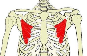
【 起 始 】:第 2(又は第 3) ~ 第5肋骨前面
 |
 |
![]()
・肩甲骨の外側(外側角周辺)を斜め下方(起始のある肋骨方向)に引く。
・肩甲骨を固定することにより第2(又は3)~第5肋骨を引き上げる。( 胸郭を広げる )
![]()
・ 神 経:資料によりばらつきが見られる。
1. 内側及び外側胸筋神経 ( C7,C8 《Th1》 ) ⇒ イラスト(一覧表5,6番)
2. 内側及び外側胸筋神経 ( C6~Th1 )
3. 内側胸筋神経 ( C7,C8 )
・ 脈 管:
 |
![]()
The pectoralis minor (/ˌpɛktəˈreɪlɪs ˈmaɪnər/) is a thin, triangular muscle, situated at the upper part of the chest, beneath the pectoralis major in the human body.
【 Structure 】
It arises from the upper margins and outer surfaces of the third, fourth, and fifth ribs, near their cartilages and from the aponeuroses covering the intercostalis.
The fibers pass superior and lateral and converge to form a flat tendon, which is inserted into the medial border and upper surface of the coracoid process of the scapula.
【Relations】
The pectoralis minor muscle is covered anteriorly (superficially) by the clavipectoral fascia. The Medial pectoral nerve pierces the pectoralis minor and the clavipectoral fascia. In attaching to the coracoid process, the pectoralis minor forms a 'bridge' - structures passing into the upper limb from the thorax will pass directly underneath.[1]
Axillary nodes are classified according to their positions relative to the pectoralis minor muscle. Level 1 are lateral, Level 2 are deep, Level 3 are medial. The pectoralis minor divides the axillary artery into three parts (in contrary sequence compared to the nodes) - first part medial, second part deep/posterior, third part lateral in relation to the pectoralis minor.
【Variations】
The origin is from the second, third and fourth or fifth ribs. The tendon of insertion may extend over the coracoid process to the greater tubercle. It may be split into several parts. Absence of this muscle is rare but happens with certain uncommon diseases, such as the Poland syndrome.[citation needed]
【 Function 】
The pectoralis minor depresses the point of the shoulder, drawing the scapula superior, towards the thorax, and throwing its inferior angle posteriorly.
【 語 句 】
・pectoralis major:大胸筋 ・cartilages:軟骨 ・aponeuroses:腱膜 ・intercostalis:肋間筋 ・coracoid process:烏口突起 ・scapula:肩甲骨 ・clavipectoral fascia:鎖骨胸筋筋膜 ・ Medial pectoral nerve:内側胸筋神経 ・Axillary nodes:腋窩リンパ節 ・greater tubercle:大結節 ・Poland syndrome:ポーランド症候群 ・thorax:胸郭
![]()
![]()