

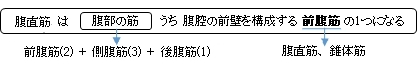
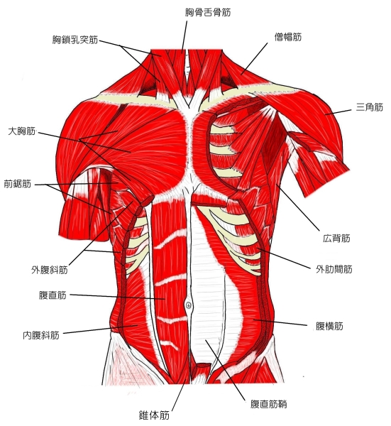
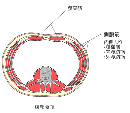
腹直筋 ( ふくちょくきん、 英 : rectus abdominis muscle )
・ 概 要 |
・ 作 用 |
・ イラスト掲載サイト |
|
・ イラスト |
・ 神経 / 脈管 |
||
・ 起始 / 停止 |
・ Wikipedia |
![]()
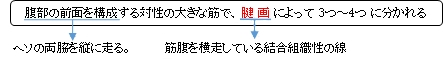
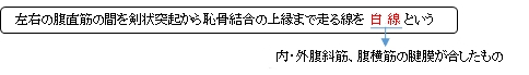
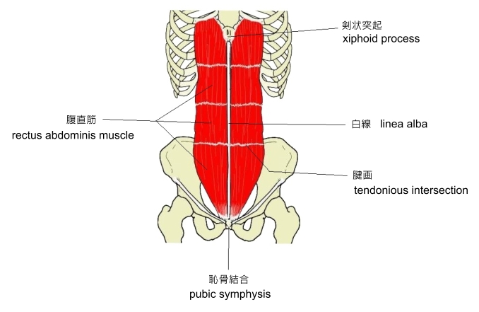
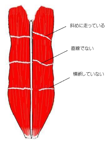
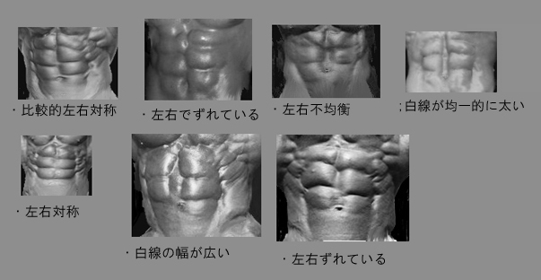
・ 腱画によって腹直筋が分かれることによって、いわゆる「シックスパック」が形作られる。
![]()
・ 「 It is typically around 10 mm thick. Although, some athletes can have a rectus up to 20 mm thick. 」 ( Wikipedia )
以下は 「 船戸和也のHP 」の解説の一部となる。
「 断面が楕円形のこの筋の停止腱から分かれた線維は、正中線を越え、白線の尾側への続きとして恥骨結合から陰茎(陰核)の背側面に向かう陰茎(陰核)提靱帯に加わる。腹直筋が強く働くのは背臥位から状態を起こすとき、ボートを漕ぐときなどである。腹筋の発達した人では腱画の位置が皮膚の上からくぼんで見える。 」
⇒ 「Rauber-Kopsch解剖学」の腹直筋の解説の「変異」の解説の引用ページへ
⇒ 「Rauber-Kopsch解剖学」の「側腹の筋群」の一覧
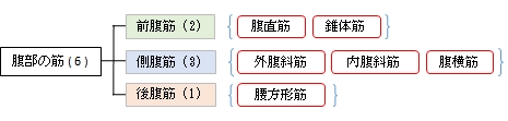
筋連結 : 大胸筋、 外腹斜筋、 内腹斜筋、 腹横筋、 錐体筋
![]()
 |
 |
 |
 |
 |
 |
![]()
【 停 止 】 : 第5~第7肋軟骨、 剣状突起 (胸骨)、 肋剣靭帯
![]()
「 腹部の圧縮、腹部内臓の保護、強い呼気時に働く、骨盤と脊柱の屈曲。」 ( 船戸和也のHP )
![]()
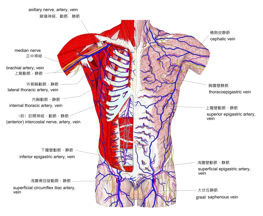
・ 脈 管 : 動脈 ) 上・下腹壁動脈の筋枝 ( 船戸和也のHP )
※ 上記の2つの動脈に加え、Wikipedia では 「下位6つの肋間動脈からの多くの枝」 を挙げている。
胸部~腹部の血管 |
|
![]()
The rectus abdominis muscle, also known as the "abdominal muscle", is a paired muscle running vertically on each side of the anterior wall of the human abdomen, as well as that of some other mammals. There are two parallel muscles, separated by a midline band of connective tissue called the linea alba. It extends from the pubic symphysis, pubic crest and pubic tubercle inferiorly, to the xiphoid process and costal cartilages of ribs V to VII superiorly. The proximal attachments are the pubic crest and the pubic symphysis. It attaches distally at the costal cartilages of ribs 5-7 and the xiphoid process of the sternum.
The rectus abdominis muscle is contained in the rectus sheath, which consists of the aponeuroses of the lateral abdominal muscles. Bands of connective tissue called the tendinous intersections traverse the rectus abdominis, which separates this parallel muscle into distinct muscle bellies. The outer, most lateral line, defining the "abs" is the linea semilunaris. In the abdomens of people with low body fat, these muscle bellies can be viewed externally and are commonly referred to as "four", "six", "eight", or "ten packs", depending on how many are visible; although, six is the most common.
【 語 句 】
・ abdomen : 腹部 ・ connective tissue : 結合組織 ・ linea alba : 白線 ・ pubic symphysis : 恥骨結合 ・ pubic crest : 恥骨稜 ・ pubic tubercle : 恥骨結節 ・ xiphoid process : 剣状突起 ・ costal cartilages : 肋軟骨 ・ proximal : 近位の ・ sternum : 胸骨 ・ rectus sheath : 腹直筋鞘 ・aponeuroses : 腱膜 ・ tendinous intersections : 腱画 ・ distinct : 明瞭な ・ linea semilunaris : 半月線 ・ externally : 外面的に
The rectus abdominis is a long flat muscle, which extends along the whole length of the front of the abdomen, and is separated from its fellow of the opposite side by the linea alba. Tendinous intersections (intersectiones tendineae) further subdivide each rectus abdominis muscle into a series of smaller muscle bellies. Tensing of the rectus abdominis causes the muscle to expand between each tendinous intersection.[3]
The upper portion, attached principally to the cartilage of the fifth rib, usually has some fibers of insertion into the anterior extremity of the rib itself.
【 Size 】
It is typically around 10 mm thick.[4] Although, some athletes can have a rectus up to 20 mm thick.[5] Typical volume is around 300 cm³ in non-active individuals and 500 cm³ in athletes.[6]
【 Blood supply 】
The rectus abdominis has many sources of arterial blood supply. Classification of the vascular anatomy of muscles: First, the inferior epigastric artery and vein (or veins) run superiorly on the posterior surface of the rectus abdominis, enter the rectus fascia at the arcuate line, and serve the lower part of the muscle. Second, the superior epigastric artery, a terminal branch of the internal thoracic artery, supplies blood to the upper portion. Finally, numerous small segmental contributions come from the lower six intercostal arteries as well.
【 語 句 】
・ principally : 主として ・ extremity : 先端 ・ arterial : 動脈の ・ Classification : 分類 ・ vascular : 血管の ・ inferior epigastric artery :下腹壁動脈 ・ rectus fascia : 腹直筋膜? ・ arcuate line : 弓状線 ・ superior epigastric artery : 上腹壁動脈 ・ internal thoracic artery : 内胸動脈 ・ intercostal arteries : 肋間動脈
【 Nerve supply 】
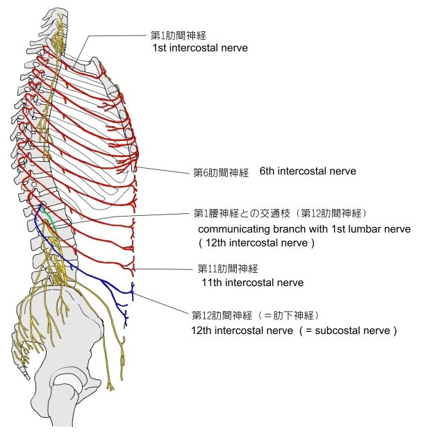
The muscles are innervated by thoraco-abdominal nerves, these are continuations of the T7-T11 intercostal nerves and pierce the anterior layer of the rectus sheath. Sensory Variation supply is from the 7-12 thoracic nerves
【 Variation 】
The sternalis muscle may be a variant form of the pectoralis major or the rectus abdominis. Some fibers are occasionally connected with the costoxiphoid ligaments, and the side of the xiphoid process.
【 Function 】
The rectus abdominis is an important postural muscle. It is responsible for flexing the lumbar spine, as when doing a so-called "crunch" sit up. The rib cage is brought up to where the pelvis is when the pelvis is fixed, or the pelvis can be brought towards the rib cage (posterior pelvic tilt) when the rib cage is fixed, such as in a leg-hip raise. The two can also be brought together simultaneously when neither is fixed in space.
The rectus abdominis assists with breathing and plays an important role in respiration when forcefully exhaling, as seen after exercise as well as in conditions where exhalation is difficult such as emphysema. It also helps in keeping the internal organs intact and in creating intra-abdominal pressure, such as when exercising or lifting heavy weights, during forceful defecation or parturition (childbirth).
【 語 句 】
・ thoraco-abdominal nerves : 胸腹神経 ・ intercostal nerves : 肋間神経 ・ thoracic nerves : 胸神経 ・ sternalis muscle : 胸骨筋 ・ variant : 異なる ・ costoxiphoid ligaments : 肋剣靭帯 ・ lumbar spine : 腰椎 ・ pelvis : 骨盤 ・ simultaneously : 同時に ・ respiration : 呼吸 ・ exhale : 息を吐きだす ・ emphysema : 肺気腫 ・ keeping the internal organs intact : 内臓を保全すること
![]()
![]()