【概 要】


【通過するもの】
以下は「ChatGPT」を参考に横行結腸間膜を通る動脈や神経などを一覧にしたものとなる。
動
脈 |
小腸への主要な動脈である 上腸間膜動脈は以下の枝を腸間膜内で分岐 |
1 |
|
|
2 |
|
|
3 |
回腸動脈 |
|
4 |
回盲動脈 |
|
5 |
|
|
6 |
|
|
静脈 |
上腸間膜静脈は
小腸からの血液を集め、脾静脈と合流して門脈を形成します。 |
神
経 |
1 |
自律神経 |
上腸間膜神経叢から分布
交感神経:内臓神経(主に胸内臓神経)を介して来る → 蠕動抑制・血管収縮など
副交感神経:迷走神経由来 → 蠕動促進・分泌促進など |
2 |
知覚神経 |
腹部の痛み(特に内臓痛)などに関与 |
3 |
|
|
| 他 |
|
|
腸間膜リンパ節、乳び槽 |
【参考となるサイト】
「日本人体解剖学」では以下のように解説している。
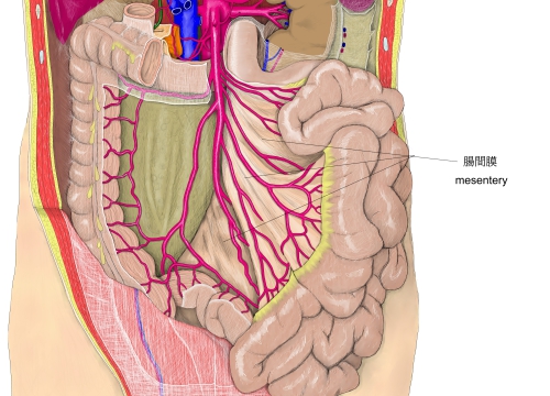
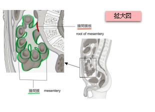
第2腰椎の左側から右腸骨窩に至る約15㎝の長さをもって壁側腹膜から起こり、ここを腸間膜根という。腸間膜は腸間膜根から長付着部に至るに従い次第にその広さを増し、空・回腸に付着する縁は数mとなる。また、腸間膜根と腸管との距離は約20㎝である。腸間膜根は、2枚の腹膜とその間に介在する疎性結合組織からなり、結合組織中には小腸に分布する血管、神経、リンパ管、リンパ節および脂肪塊が含まれる。
以下は「Wikipedia」の解説文となる。 mesentery
In human anatomy, the mesentery is an organ that attaches the intestines to the posterior abdominal wall, consisting of a double fold of the peritoneum. It helps (among other functions) in storing fat and allowing blood vessels, lymphatics, and nerves to supply the intestines.[1]
The mesocolon (the part of the mesentery that attaches the colon to the abdominal wall) was formerly thought to be a fragmented structure, with all named parts—the ascending, transverse, descending, and sigmoid mesocolons, the mesoappendix, and the mesorectum—separately terminating their insertion into the posterior abdominal wall.[2] However, in 2012, new microscopic and electron microscopic examinations showed the mesocolon to be a single structure derived from the duodenojejunal flexure and extending to the distal mesorectal layer.[2][3] Thus the mesentery is an internal organ.[4][5]
Structure
The mesentery of the small intestine arises from the root of the mesentery (or mesenteric root) and is the part connected with the structures in front of the vertebral column. The root is narrow, about 15 cm long, 20 cm in width, and is directed obliquely from the duodenojejunal flexure at the left side of the second lumbar vertebra to the right sacroiliac joint. The root of the mesentery extends from the duodenojejunal flexure to the ileocaecal junction. This section of the small intestine is located centrally in the abdominal cavity and lies behind the transverse colon and the greater omentum.
The mesentery becomes attached to the colon at the gastrointestinal margin and continues as the several regions of the mesocolon. The parts of the mesocolon take their names from the part of the colon to which they attach. These are the transverse mesocolon attaching to the transverse colon, the sigmoid mesocolon attaching to the sigmoid colon, the mesoappendix attaching to the appendix, and the mesorectum attaching to the upper third of the rectum.
The mesocolon regions were traditionally taught to be separate sections with separate insertions into the posterior abdominal wall. In 2012, the first detailed observational and histological studies of the mesocolon were undertaken and this revealed several new findings.[6] The study included 109 patients undergoing open, elective, total abdominal colectomy. Anatomical observations were recorded during the surgery and on the post-operative specimens.
These studies showed that the mesocolon is continuous from the ileocaecal to the rectosigmoid level. It was also shown that a mesenteric confluence occurs at the ileocaecal and rectosigmoid junctions, as well as at the hepatic and splenic flexures and that each confluence involves peritoneal and omental attachments. The proximal rectum was shown to originate at the confluence of the mesorectum and mesosigmoid. A plane occupied by perinephric fascia was shown to separate the entire apposed small intestinal mesentery and the mesocolon from the retroperitoneum. Deep in the pelvis, this fascia coalesces to give rise to presacral fascia.[6]
【 語 句 】
・: ・: ・: ・: ・: ・: ・: ・: ・: ・: ・: ・: ・: ・: ・: ・: ・: ・: ・: ・: ・: ・: ・: ・: ・: ・: ・: ・: ・: ・: ・: ・:
■ 写真やイラストを掲載しているサイト ■
・ イラストや写真を掲載しているサイト-Ⅰ
・ イラストや写真を掲載しているサイト-Ⅱ
・ イラストや写真を掲載しているサイト-Ⅲ
・ イラストや写真を掲載しているサイト-Ⅳ
・ イラストや写真を掲載しているサイト-Ⅴ