

広背筋 (こうはいきん、英:latissimus dorsi muscle)
・ 概 要 |
・ 作 用 |
・ イラスト掲載サイ |
|
・ イラスト |
・ 神経 / 脈管 |
||
・ 起始 / 停止 |
・ Wikipedia |
![]()
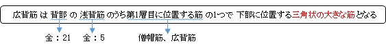
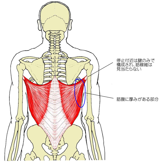
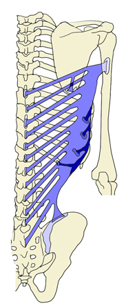
・ 比較的に薄い筋だが、上外側方の部位には厚みが見られる。
・ 上部内側方の部分は 僧帽筋 に覆われる。
・ 腕を挙上した場合、広背筋の上外側部は身体の前面からでも確認ができる。
【 変 異 】
「 船戸和弥のHP 」 の 「 Rauber-Kopsch解剖学 」 ではこの筋の解説において「変異」ということで以下のように解説している。
「 変 異 : 個々の筋束に分離することがある.棘突起,腸骨稜および肋骨における筋起始の欠如あるいは増加がありうる(肋骨からの起始部についてはFrey, Vierteljahrsschr. naturforsch, Ges. Zürich 1918参照).腰三角の形は,この筋が腸骨稜から起るぐあいによって異る.肩甲骨下角から出る副筋束は,大体いつも見られる.広背筋停止腱は,大円筋の腱と分けられないことが多い.過剰筋束としては,広背筋の外側縁において非常に不定であるが腱性あるいは筋性の索が 7~8% (W. Krause, Le Double) に見られる.これは広背笏の外側縁から分れてでて,腋窩を超えて,多くは大胸筋腱の後面に終るが,また他の場所にも停止することがある.この筋束に対してランゲル腋窩弓 “Langer-scher Achselbogen” という名称を用いる.
他の副筋束としては,長肘筋M. anconaeus longus(Henle),または広背顆筋M. latissimocondyloideus(Bischoff)とよばれるものがある.これは約5%に見られ,広背筋の腱または筋腹に始まり,上腕筋膜,上腕三頭筋,上腕骨に終り,なお遠く前腕の筋膜にも停止することがある.Ruge, G., Morph. Jahrb., 48. Bd.,1914.--Bluntshli, Morph. Jahrb., 41. Bd.,1910.
【 関連語句 】
![]()
 |
 |
 |
 |
 |
 |
![]()
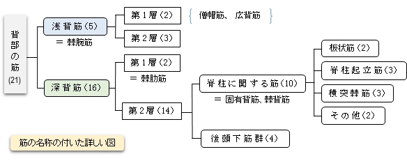
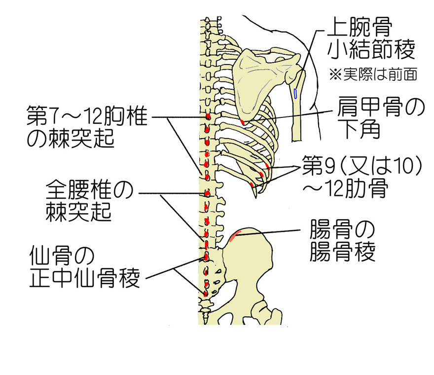
【 起 始 】 : ・胸腰筋膜浅葉 ・第7~12胸椎の棘突起 ・全腰椎の棘突起 ・仙骨の正中仙骨稜(5番)
・腸骨稜 ・第9(又は10)~12肋骨 ・肩甲骨の下角(一覧表6番)
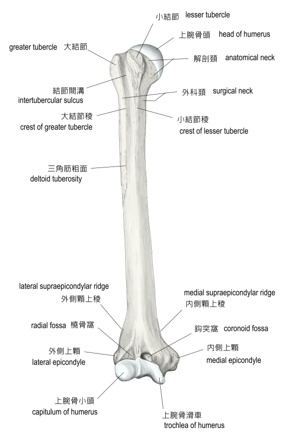
【 停 止 】 : ・上腕骨の小結節稜
 |
 |
 |
![]()
肩関節を内転、伸展、内旋させる。
![]()
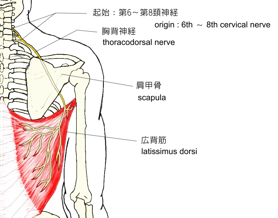
・ 神 経 : 胸背神経 ( C6,C7,C8 )
・ 脈 管 : 胸背動脈 ( 肩甲下動脈 )
 |
![]()
The latissimus dorsi (/ˌləˈtɪsɨməs ˈdɒrsaɪ/) (plural: latissimi dorsi), meaning 'broadest [muscle] of the back' (Latin latus meaning 'broad', latissimus meaning 'broadest' and dorsum meaning the back), is the larger, flat, dorso-lateral muscle on the trunk, posterior to the arm, and partly covered by the trapezius on its median dorsal region. Latissimi dorsi are commonly known as "lats", especially among bodybuilders.
The latissimus dorsi is responsible for extension, adduction, transverse extension also known as horizontal abduction, flexion from an extended position, and (medial) internal rotation of the shoulder joint. It also has a synergistic role in extension and lateral flexion of the lumbar spine.
Due to bypassing the scapulothoracic joints and attaching directly to the spine, the actions the latissimi dorsi have on moving the arms can also influence the movement of the scapulae, such as their downward rotation during a pull up.
【 語 句 】
・ trapezius : 僧帽筋 ・ adduction : 内転 ・ abduction : 外転 ・ internal rotation : 内旋? ・ synergistic : 相乗的な ・ scapulothoracic joint : ?
Contents
Structure
Variations
Axillary arches shown from two different angles.
The number of dorsal vertebrae to which it is attached varies from four to eight; the number of costal attachments varies; muscle fibers may or may not reach the crest of the ilium.
A muscular slip, the axillary arch, varying from 7 to 10 cm in length, and from 5 to 15 mm in breadth, occasionally springs from the upper edge of the latissimus dorsi about the middle of the posterior fold of the axilla, and crosses the axilla in front of the axillary vessels and nerves, to join the under surface of the tendon of the pectoralis major, the coracobrachialis, or the fascia over the biceps brachii. This axillary arch crosses the axillary artery, just above the spot usually selected for the application of a ligature, and may mislead a surgeon. It is present in about 7% of the population and may be easily recognized by the transverse direction of its fibers. Guy et al. extensively described this muscular variant using MRI data and positively correlated its presence with symptoms of neurological impingement.[1]
A fibrous slip usually passes from the upper border of the tendon of the Latissimus dorsi, near its insertion, to the long head of the triceps brachii. This is occasionally muscular, and is the representative of the dorsoepitrochlearis brachii of apes.[2][3] This muscular form is found in ~5% of humans and is sometimes termed the latissimocondyloideus.[2]
The latissimus dorsi crosses the inferior angle of the scapula. A study found that, of 100 cadavers dissected:[4]
- 43% had "a substantial amount" of muscular fibers in the latissimus dorsi originating from the scapula.
- 36% had few or no muscular fibers, but a "soft fibrous link" between the scapula and the latissimus dorsi
- 21% had little or no connecting tissue between the two structures.
【 語 句 】
・ axillary arch : 腋窩弓 ・ vertebrae : 椎骨 ・ crest of the ilium : 腸骨稜 ・ axilla : 腋窩 ・pectoralis major : 大胸筋 ・ coracobrachialis : 烏口腕筋 ・ biceps brachii : 上腕二頭筋 ・ ligature : 結紮(けつさつ)糸. ・ correlate : 互いに関係がある ・ impingement : 衝突、侵害 ・ dorsoepitrochlearis :? ・ latissimocondyloideus :? ・ cadaver : 死体 ・ dissect : 解剖する ・ substantial : しっかりした
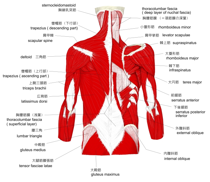
Triangles
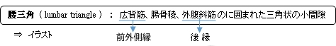
- The lateral margin of the latissimus dorsi is separated below from the obliquus externus abdominis by a small triangular interval, the lumbar triangle of Petit, the base of which is formed by the iliac crest, and its floor by the obliquus internus abdominis.
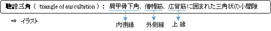
- Another triangle is situated behind the scapula. It is bounded above by the trapezius, below by the latissimus dorsi, and laterally by the vertebral border of the scapula; the floor is partly formed by the rhomboideus major. If the scapula is drawn forward by folding the arms across the chest, and the trunk bent forward, parts of the sixth and seventh ribs and the interspace between them become subcutaneous and available for auscultation. The space is therefore known as the triangle of auscultation.
- The latissimus dorsi can be remembered best for insertion as "The Lady Between Two Majors". As the latissimus dorsi inserts into the floor of the intertubercular groove of the humerus it is surrounded by two major muscles. The teres major inserts medially on the medial lip of the intertubercular groove and laterally the pectoralis major inserts into the lateral lip.
【 語 句 】
・ obliquus externus abdominis : 外腹斜筋 ・ rhomboideus major : 大菱形筋 ・ subcutaneous : 皮下の ・ auscultation : 聴診 ・ triangle of auscultation : 聴診三角 ・ intertubercular groove : 結節間溝 ・ teres major : 大円筋 ・ medial lip : 内側唇
Innervation
The latissimus dorsi is supplied by the sixth, seventh, and eighth cervical nerves through the thoracodorsal (long scapular) nerve. Electromyography suggests that it consists of six groups of muscle fibres that can be independently coordinated by the central nervous system.[5]
Function
The latissimus dorsi is responsible for extension, adduction, transverse extension also known as horizontal abduction, flexion from an extended position, and (medial) internal rotation of the shoulder joint.[citation needed] It also has a synergistic role in extension (posterior fibers) and lateral flexion (anterior fibers) of the lumbar spine, and assists as a muscle of both forced expiration (anterior fibers) and an accessory muscles of inspiration (posterior fibers).[6]
Most latissimus dorsi exercises concurrently recruit the teres major, posterior fibres of the deltoid, long head of the triceps brachii, among numerous other stabilizing muscles. Compound exercises for the 'lats' typically involve elbow flexion and tend to recruit the biceps brachii, brachialis, and brachioradialis for this function. Depending on the line of pull, the trapezius muscles can be recruited as well; horizontal pulling motions such as rows recruit both latissimus dorsi and trapezius heavily.
Trainin
The power/size/strength of this muscle can be trained with a variety of different exercises. Some of these include:
- Vertical pulling movements such as pull-downs and pull-ups (including chin-ups)
- Horizontal pulling movements such as bent-over row, T-bar row and other rowing exercises
- pull-overs
- deadlift
Lifting under control can help reduce chances of injury.
【 語 句 】
・ thoracodorsal (long scapular) nerve : 胸背神経 ・ electromyography : 筋電図検査 ・ expiration : 息を吐きだすこと ・ inspiration : 息を吸い込むこと ・ concurrently : 同時に ・ deltoid :三角筋 ・ brachialis : 上腕筋
![]()
![]()