
【経 過】
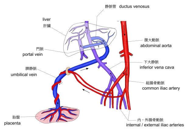
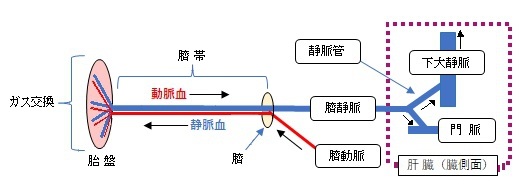
1. 臍動脈からの静脈血は胎盤でガス交換を行い、動脈血となって臍静脈へ流れていく。
2. 臍動脈とともに臍帯中を臍まで走る。
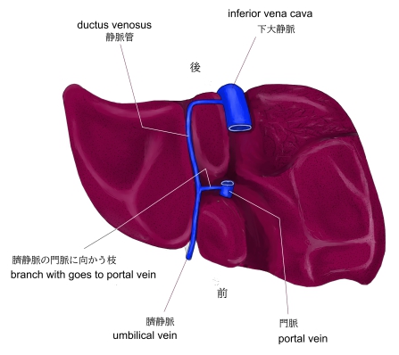
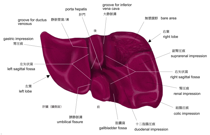
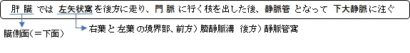
3. 臍からは肝臓の臓側面に達すると左矢状窩を後方へ走る。
4. 肝門側部で門脈に行く枝を出したのち、静脈管となって下大静脈に注ぐ。
【長 さ】:「約40〜60cm程度が一般的です。」(ChatGPT)
【流入量】
臍静脈の血液は門脈と下大静脈に注ぐが、その流入量は下大静脈へ注ぐ方が多いようである。
・「静脈管は肝臓を迂回するため、胎盤からの大部分の血液は肝臓の毛細血管網を通過することなく、直接に心臓に注ぐ。」(船戸和也のHP)
・「AI」に聞いてみたところ:「臍静脈の血流のうち、約50〜60%は静脈管を通って下大静脈へ。」
【その他】
・「通常は出生後、臍動脈が閉じてから臍静脈も閉じる形となる」(Wikipedia)
「 日本人体解剖学 」では以下のように解説している。
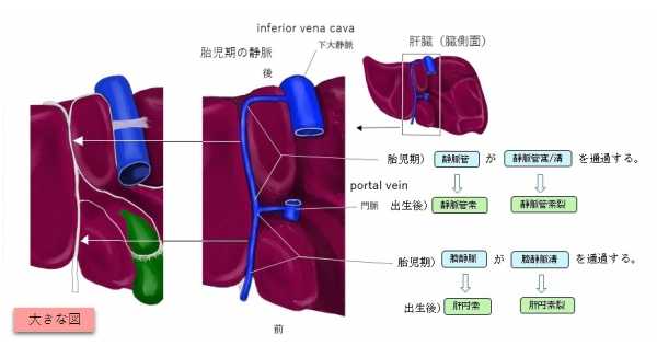
「 胎盤でガス交換し動脈血となり栄養物を与えられた血液は1本の臍静脈を通り、臍帯中を臍動脈と平行して胎児の臍輪に達する。臍輪で、臍静脈は臍動脈から離れて肝鎌状間膜中を通り肝臓の下面に達し、ここで2枝に分かれる。その1枝は静脈管で、肝臓の下面を通って下大静脈に入る。他の1枝は門脈に合流する。」
また、「 船戸和弥のHP 」では以下のように解説している。
「 臍静脈は肝臓の両側を走行し、胎盤からの酸素に富んだ血液を送るようになる。右臍静脈は胚子期の終わりに消失するため、左臍静脈だけが胎盤からの酸素に富んだ血液を胚子に送る。このような細静脈の構造的変化は以下に述べる様に要約される(図14-5)。すなわち、
・右臍静脈と肝臓と静脈洞の間に位置する臍静脈尾側部は退行する。
・左臍静脈尾側部の退行しない部位が臍静脈として存続する。
・肝臓内に大きな吻合静脈である静脈管が発生して(図14-5C)、臍静脈と下大静脈inferior vena cava(IVC)を連結する。
静脈管は肝臓を迂回するため、胎盤からの大部分の血液は肝臓の毛細血管網を通過することなく、直接に心臓に注ぐ。(Moore人体発生学)) 」
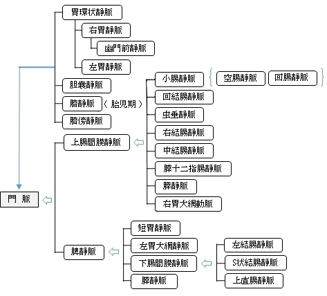
以下は門脈に注ぐ静脈を簡単に表したもとなる。
以下は「 Wikipedia 」の解説文となる。
「 The umbilical vein is a vein present during fetal development that carries oxygenated blood from the placenta into the growing fetus. The umbilical vein provides convenient access to the central circulation of a neonate for restoration of blood volume and for administration of glucose and drugs.[1]
The blood pressure inside the umbilical vein is approximately 20 mmHg.[2]
【 Fetal circulation 】
The unpaired umbilical vein carries oxygen and nutrient rich blood derived from fetal-maternal blood exchange at the chorionic villi. More than two-thirds of fetal hepatic circulation is via the main portal vein, while the remainder is shunted from the left portal vein via the ductus venosus to the inferior vena cava, eventually being delivered to the fetal right atrium.
【 語 句 】
・ fetal development : 胎児発生 ・ oxygenated : 酸素量が増した ・ placenta : 胎盤 ・fetus : 胎児 ・ neonate : 新生児 ・ restoration : 元に戻すこと ・ administration : 管理 ・ glucose : ぶどう糖 ・ chorionic villi : 絨毛膜絨毛 ・ portal vein : 門脈 ・ ductus venosus : 静脈管 ・ inferior vena cava : 下大静脈 ・ right atrium : 右心房
【 Closure 】
Closure of the umbilical vein usually occurs after the umbilical arteries have closed. This prolongs the communication between the placenta and fetal heart, allowing for a sort of autotransfusion of remaining blood from the placenta to the fetus.
Within a week of birth, the neonate's umbilical vein is completely obliterated and is replaced by a fibrous cord called the round ligament of the liver (also called ligamentum teres hepatis). It extends from the umbilicus to the transverse fissure, where it joins with the falciform ligament of the liver to separate segment 4 from segments 2 and 3 of the left hepatic lobe.
【 語 句 】
・ prolong : 延長する ・ autotransfusion : 自己輸血 ・ obliterate : 抹消する ・ round ligament of the liver / ligamentum teres hepatis : 肝円索 ・ umbilicus : へそ ・ fissure : 裂孔 ・ falciform ligament : かま状靭帯 ・ hepatic lobe : 肝葉
【 Recanalization 】
Under extreme pressure, the round ligament may reopen to allow the passage of blood. Such recanalization may be evident in patients with cirrhosis and portal hypertension. Patients with cirrhosis experience rapid growth of scar tissue in and around the liver, often functionally obstructing nearby vessels. Vessel occlusion increases vascular resistance and therefore leads to hypertension. In portal hypertension, the vessels surrounding the liver are subjected to abnormally high blood pressure—so high, in fact, that the force of the blood pressing against the round ligament is sufficient to recanalize the structure. This leads to a condition called Caput medusae.
【 Catheterization 】
A newborn baby has a patent umbilical vein for at least a week after birth. This umbilical vein may be catheterised for ready intravenous access. It may be used as a site for regular transfusion in cases of erythroblastosis or hemolytic disease. It also provides a route for measuring central venous pressure.[1] 」
【 語 句 】
・ Recanalization : 血流再開 ・ evident : 明白な ・ cirrhosis : 肝硬変 ・ portal hypertension : 門脈圧亢進症 ・ scar tissue : 瘢痕(はんこん)組織 ・ occlusion : 閉塞 ・ recanalize : 再開通する ・ Caput medusae : メデューサの頭 ・ patent : 明白な? ・ catheterise ~ : ~ にカテーテルを挿入する ・ ntravenous : 静脈内の ・ erythroblastosis : 赤芽球症 ・ hemolytic : 溶血性の
【 イラスト掲載サイト 】
・ イラストや写真を掲載しているサイト-Ⅰ
・ イラストや写真を掲載しているサイト-Ⅱ
・ イラストや写真を掲載しているサイト-Ⅲ

|