恥骨筋 ( ちこつきん、英:pectineus muscle )
・ 概 要 |
・ 作 用 |
・ イラスト掲載サイト |
|
・ イラスト |
・ 神経 / 脈管 |
||
・ 起始 / 停止 |
・ Wikipedia |
![]()


![]()
以下は「船戸和也のHP」の解説文となる。
「恥骨筋は腸恥隆起-恥骨結節間の恥骨上肢から起こり、大腿骨の恥骨筋線に停止する。この筋はもとももと腸腰筋群と同じ原基に由来する。本筋の構成に内転筋群がどの程度関わるかには個体差がある。恥骨筋は腸骨筋膜の延長部分である恥骨筋膜におおわれ、腸腰筋とともに腸恥窩には大腿動静脈が通る。」
![]()
 |
 |
 |
![]()
【 起 始 】
【 停 止 】 : 恥骨筋線(大腿骨)
![]()
「大腿を内転し、屈する。 」 ( 日本人体解剖学 )
※「大腿=股関節」
![]()
 |
![]()
Pectineus muscle is a flat, quadrangular muscle, situated at the anterior part of the upper and medial aspect of the thigh. The pectineus muscle is the most anterior dductor of the hip .
It can be classified in the medial compartment of thigh (when the function is emphasized) or the anterior compartment of thigh (when the nerve is emphasized).
【Origin】
It has the most superior attachment of all the thigh adductors, originating from the pectineal line of pubis on the superior pubic ramus. The muscle then slides over the superior margin of superior pubic ramus and courses posterolaterally down the thigh, sometimes being partially divided into a larger anterior (superficial) layer and smaller posterior (deep) layer. The layers are innervated by different nerves.
【Insetion】
Pectineus muscle inserts into the posterior surface of femur, along the pectineal line and proximal part of linea aspera.
【Nerve supply】
Pectineus is predominately innervated by the femoral nerve (L2, L3)]. However, in some people pectineus may receive innervation from two separate nerves of the lumber plexus.
This composite innervation reflects the dual compartmentalisation of pectineus into both the anterior and medial compartments of the thigh. In these cases the anterior part of the muscle sits is innervated by the femoral nerve (L2, L3), a feature of muscles of the anterior thigh. While the posterior, smaller part of the muscle is supplied by a branch of obturator nerve (L2, L3), the accessory obturator nerve.
【 語 句 】
・quadrangular:四辺形の ・pectineal line:恥骨筋腺 ・pubis:恥骨 ・superior pubic ramus:恥骨上枝 ・proximal:近位の ・linea aspera:粗線 ・predominately:優位に ・femoral nerve:大腿神経 ・lumber plexus:腰神経叢 ・obturator nerve:閉鎖神経
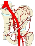
【Blood supply】
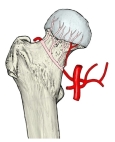
The superficial part of the muscle is supplied by the medial circumflex femoral artery, a branch of the femoral artery. Deep portion of the muscle is vascularised by the anterior branch of obturator artery, itself a branch of the internal iliac artery.
【Relation】
The muscle lies in the frontal plane and medially to,adductor longus. While laterally, it is related to the psoas major muscle and the medial circumflex femoral artery and vein.
The anterior surface of pectineus forms the medial part of the floor of femoral traingle together with adductor longus.
This surface of pectineus is covered with the deep layer of fascia lata that separates it from the femoral artery, femoral vein and great saphenous vein that course through the femoral triangle.
Posterior to pectineus are the adductor magnus, adductor brevis and obturator externus muscles, and the anterior branch of obturator nerve.
【Action】
Due to the course of its fibers, pectineus both flexes and adducts the thigh at the hip joint when it contracts. When the lower limb is in the anatomical position, contraction of the muscle first causes flexion to occur at the hip joint. This flexion can go as far as the thigh being at a 45 degree angle to the hip joint.
At that point, the angulation of the fibers is such that the contracted muscle fibers now pull the thigh towards the midline, producing thigh adduction.
【 語 句 】
・medial circumflex femoral artery:内側大腿回旋動脈 ・femoral artery:大腿動脈 ・vascularise:血管を作る ・obturator artery:閉鎖動脈 ・ internal iliac artery:内腸骨動脈 ・adductor longus:長内転筋 ・psoas major:大腰筋 ・femoral triangle:大腿三角 ・fascia lata: ・angulationgreat saphenous vein: ・adductor magnus: ・adductor brevis: ・:obturator externus ・contract: ・angulation:
![]()


