

膝蓋骨 ( しつがいこつ、英:patella )
|
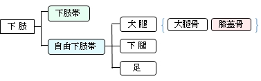
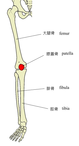
・膝蓋骨は自由下支帯の大腿の骨の一つで、「日本人体解剖学 (上巻)
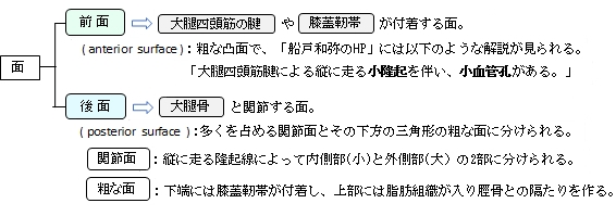
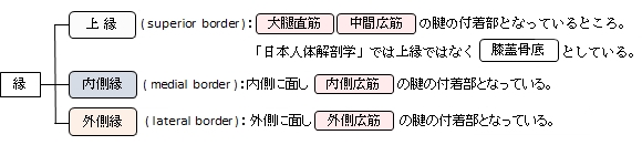
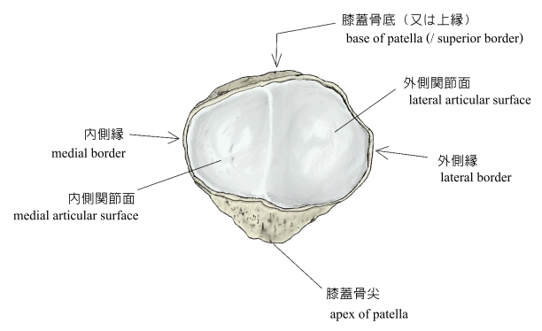
・栗の実に似た三角形の扁平な骨で以下のような特徴がある。 【前面と後面の2面を有する】 【上縁、内側縁、外側縁の3縁の区別ができる】 「日本人体解剖学 (上巻)
・以下の文章は「日本人体解剖学」(南山堂)からの引用文となる。 「発生:骨化は3歳ころに始まり、20歳ころに完了する。」 「異常:欠けることがある。上・下の2部に分かれ、その間に靭帯の入ることがある。」 ⇒「Variations in Shapes of Patella Partita」というタイトルでイラストを掲載しているサイト (※「partita」(音楽用語のこと?)とは「partite」(深裂)とのことなのだろうか?) ・「Wikipedia」の「patella」の解説ページの「Variation」には以下のような解説が見られる。 「Emarginations (i.e. patella emarginata, a "missing piece") are common laterally on the proximal edge.[1] Bipartite patellas are the result of an ossification of a second cartilaginous layer at the location of an emargination. Previously, bipartite patellas were explained as the failure of several ossification centres to fuse, but this idea has been rejected. Partite patellas occur almost exclusively in men. Tripartite and even multipartite patellas occur.」 ⇒「Classification of Biparitie Patella」というタイトルでイラストやレントゲン画像を掲載しているサイト ⇒「BIPARTITE PATELLA」というタイトルのイラストを掲載しているサイト ⇒ bipartiteのタイプを3通りに分けてイラストを掲載しているサイト
|

![]()
|
|
 |
 |
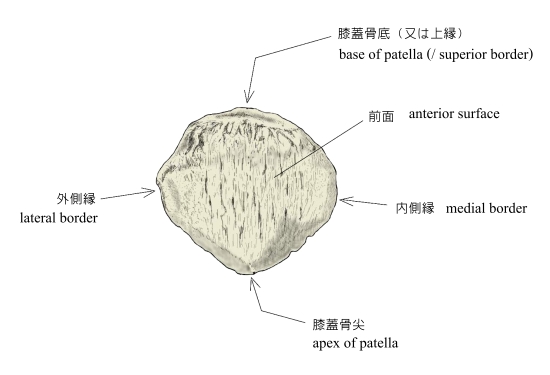
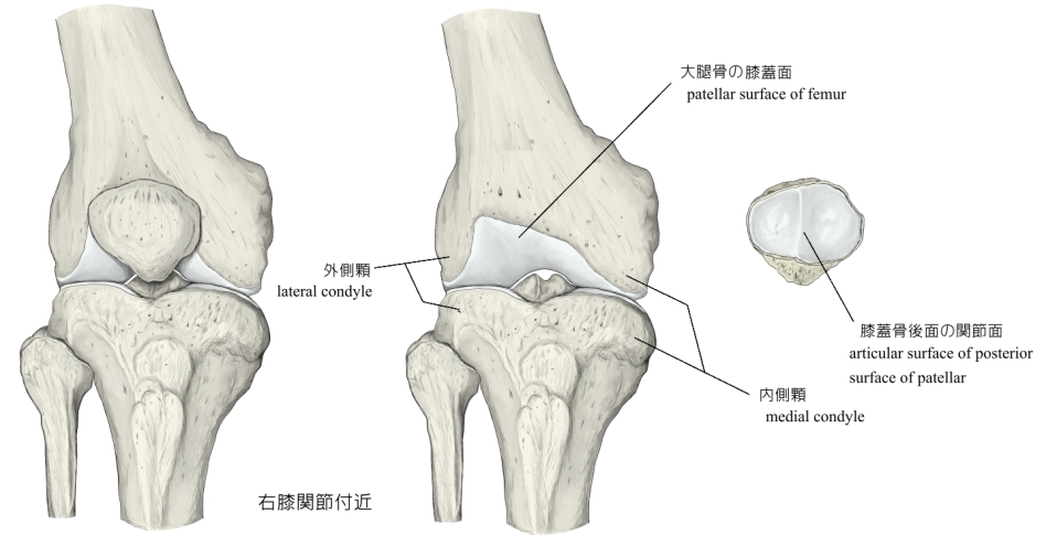
右膝蓋骨(前面) |
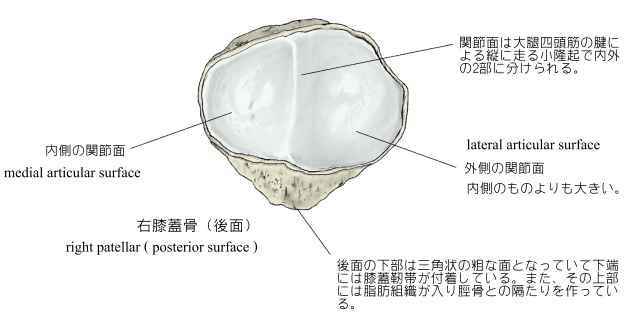
右膝蓋骨(後面) |
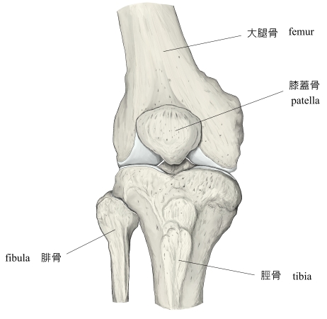
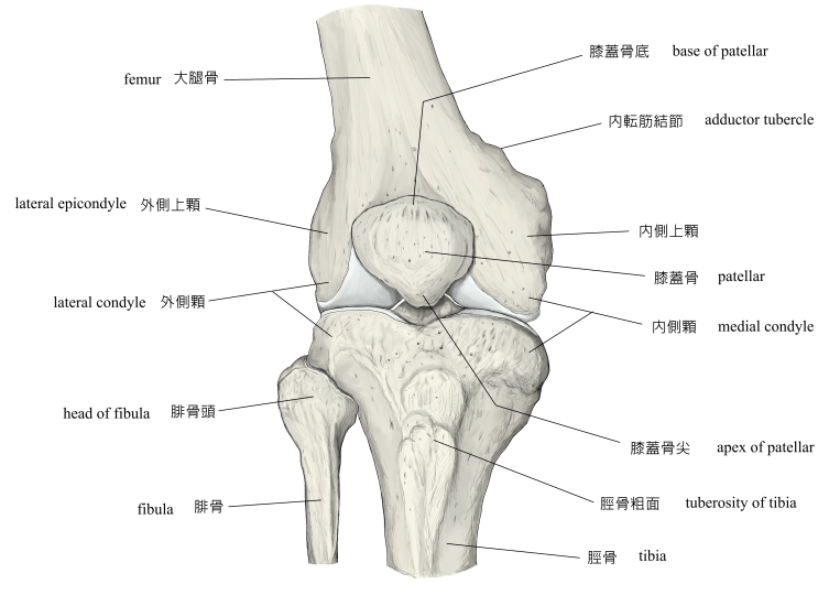
右膝関節周辺(前面) |
右膝関節周辺(矢状断面 |
⇒ レントゲン画像を掲載しているサイト-Ⅰ(膝関節を側方より)
⇒ レントゲン画像を掲載しているサイト-Ⅱ(膝蓋骨と大腿骨滑車)

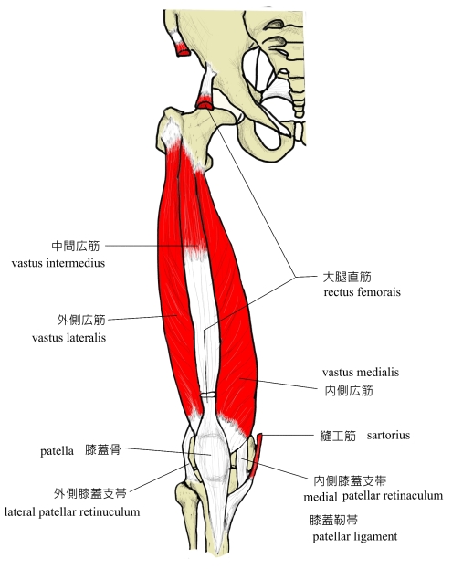

以下が膝蓋骨に付着する靭帯となる。
1 |
・「Wikipedia」の「patella」の解説ページの「Muscles」には以下のような解説が見られる。
「The patella is stabilized by the insertion of the horizontal fibres of vastus medialis and by the prominence of the lateral femoral condyle, which discourages lateral dislocation during flexion. The retinacular fibres of the patella also stabilize it during exercise.」
![]()
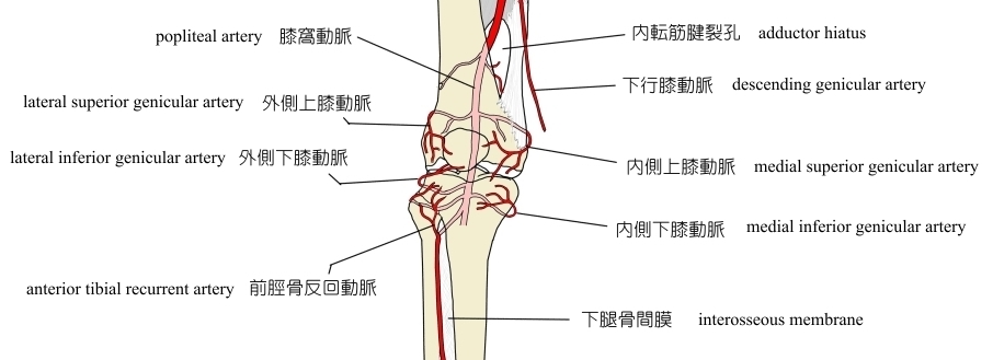
膝蓋骨へ血液を供給している動脈の名称を挙げているサイトは見つからないのだが、おそらく以下の2つの動脈がメインになるのではないだろうか。(※他にもある可能性もあるし、正確性に欠ける可能性もありうる)
|
以下のように解説しているサイトもある。
「into patella;
- recurrent branch of anterior tibial artery;
- emerges from anterior tibial artery as it perforates interosseous foramen;
- this branch passes superiorly and in the direction of patellar tendon, supplying the skin overlying patellar tendon;
- descending branch of the lateral femoral circumflex artery;
- one of the major communications between deep and superficial femoral systems of the leg;
- it travels down fascia lata & sends multiple perforators to skin overlying fascia lata & lateral aspect of knee;」
![]()
・SKELETAL SYSTEM ANATOMY: Bones of the lower extremity- Patella (1:59)
・Anatomy Of The Patellar Tendon - Everything You Need To Know - Dr. Nabil Ebraheim (5:21)