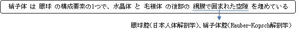
・「前後から少し圧平された球の形をしている」(Rauber-Kopsch解剖学)
・「It makes up four-fifths of the volume of the eyeball.」(Wikipedia)


|
 |
 |
 |
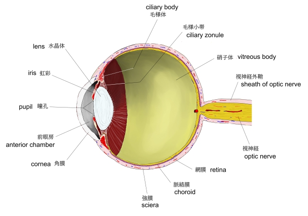
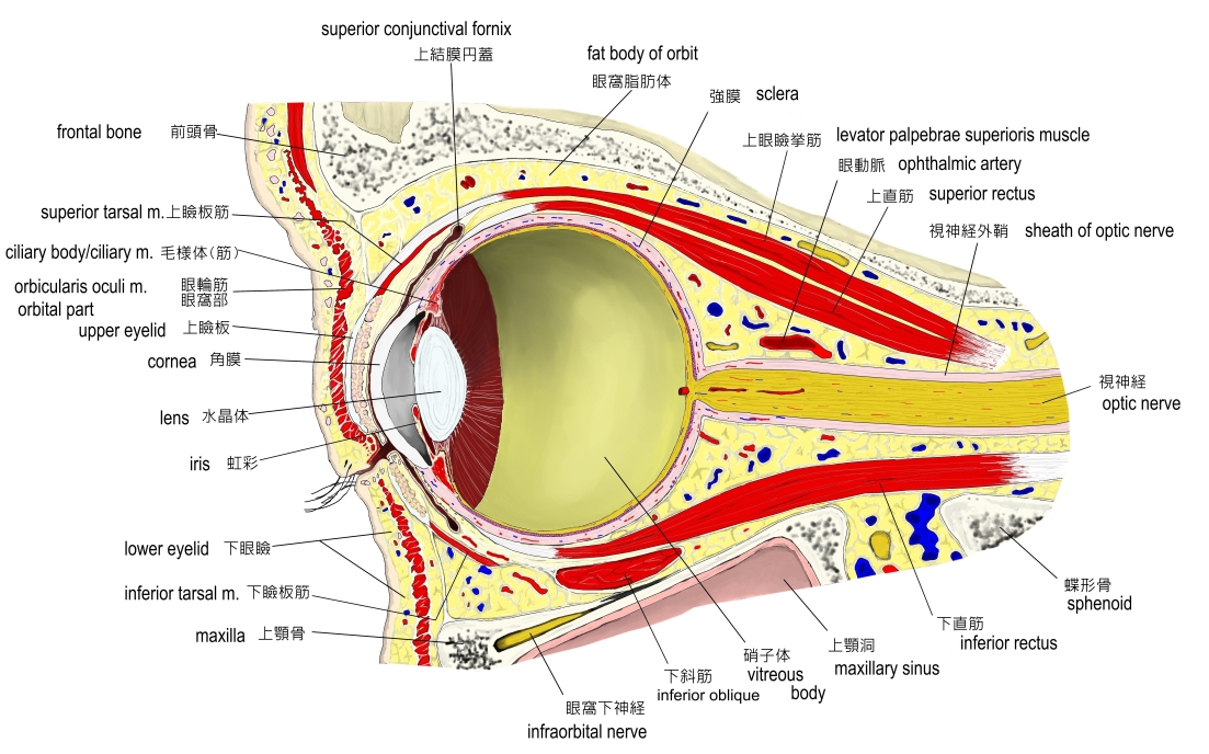
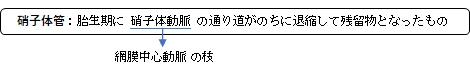
眼球(縦断面) |
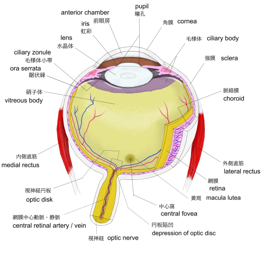
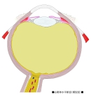
眼球・横断面-1 |
眼球・横断面-1 |
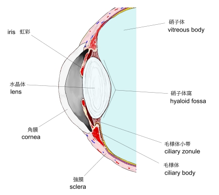
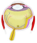
眼球前面・縦断面 |
 |
|
|
|
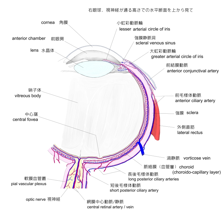
| 眼球の血管 |
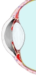
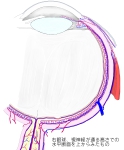
眼窩部(縦断面) |
|
|


以下は「Wikipedia」の解説文となる。
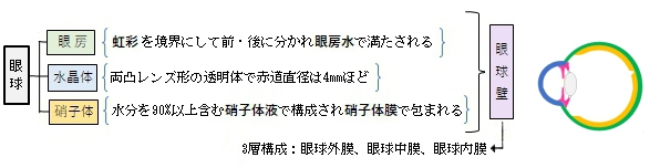
The vitreous body (vitreous meaning "glass-like"; from Latin vitreus 'glassy', from vitrum 'glass', and -eus) is the clear gel that fills the space between the lens and the retina of the eyeball (the vitreous chamber) in humans and other vertebrates. It is often referred to as the vitreous humor (also spelled humour, from Latin meaning liquid) or simply "the vitreous". Vitreous fluid or "liquid vitreous" is the liquid component of the vitreous gel, found after a vitreous detachment. It is not to be confused with the aqueous humor, the other fluid in the eye that is found between the cornea and lens.
【 語 句 】
・lens:水晶体 ・retina:網膜 ・vitreous chamber:硝子体腔 ・vertebrate:脊椎動物 ・vitreous humor:硝子体液 ・vitreous detachment:硝子体剥離 ・aqueous humor:眼房水 ・cornea:角膜
【Structure 】
The vitreous humor is a transparent, colorless, gelatinous mass that fills the space in the eye between the lens and the retina. It is surrounded by a layer of collagen called the vitreous membrane (or hyaloid membrane or vitreous cortex) separating it from the rest of the eye. It makes up four-fifths of the volume of the eyeball.The vitreous humour is fluid-like near the centre, and gel-like near the edges.
The vitreous humour is in contact with the vitreous membrane overlying the retina. Collagen fibrils attach the vitreous at the optic nerve disc and the ora serrata (where the retina ends anteriorly), at the Wieger-band, the dorsal side of the lens. The vitreous also firmly attaches to the lens capsule, retinal vessels, and the macula, the area of the retina which provides finer detail and central vision.
Aquaporin 4 in Müller cells in rats transports water to the vitreous body.
【 語 句 】
・transparent:透明な ・gelatinous:ゼラチン状の ・vitreous membrane:硝子体膜 ・hyaloid:透明な ・vitreous cortex:硝子体支質 ・fibril:原繊維 ・optic nerve disc:視神経円板 ・ora serrata:鋸状縁 ・dorsal: ・lens capsule:水晶体包 ・macula:網膜黄斑 ・aquaporin:アクラポリン(タンパク質の一種) ・Müller cells:ミューラー細胞
Anatomical features
The vitreous has many anatomical landmarks, including the hyaloid membrane, Berger's space, space of Erggelet, Wieger's ligament, Cloquet's canal and the space of Martegiani.
Surface features:
- Patella fossa: Shallow saucer-like concavity anteriorly, in which the lens rests, separated by Berger's space
- Ligamentum hyaloideocapsulare (Wieger's ligament): Circular thickening of vitreous 8-9mm in diameter, delineates the patella fossa
- Anterior hyaloid: Vitreous surface anterior to ora serrata. Continuous with and invests in the zonular fibres, and extends forward between the ciliary processes
- Vitreous base: Denser cortical area of vitreous. Firmly attached to the posterior 2mm of the pars plana, and the anterior 2-4mm of retina
- Posterior hyaloid surface: Closely applied to retinal internal limiting membrane. Firm attachment sites: Along blood vessels and at sites of retinal degeneration
- Space of Martegioni: A funnel shaped space overlying the optic disc with condensed edge
- Cloquet's canal: A 1–2 mm wide canal within the vitreous, from the space of Martegioni to the space of Berger, along an S-shaped course mainly below the horizontal.
- Mittendorf's dot: A small circular opacity on the posterior lens capsule, which represents the site of attachment of the hyaloid artery before it subsequently regressed.[8]
- Bergmeister's papilla: A tuft of fibrous tissue at the optic disc, which represents a remnant of the sheath associated with the hyaloid artery before it subsequently regressed.
【 語 句 】
・: ・: ・: ・: ・: ・: ・: ・: ・: ・: ・: ・: ・: ・: ・: ・: ・: ・: ・: ・: ・: ・: ・: ・: ・: ・: ・: ・: ・: ・: ・: ・: ・: ・: ・: ・:
internal structures of the vitreous
- The vitreous body at birth is homogenous with a finely striated pattern.
- With early aging the vitreous develops narrow transvitreal "channels".
- The cortex is denser than the centre with development.
- From adolescence, vitreous tracts form from anterior to posterior.
- These vitreous tracts are fine sheet-like condensations of vitreous.
Named tracts
- Retrolental tract: Extends posteriorly from the hyaloideocapsular ligament into central vitreous
- Coronary tract: External to the retrolental tract, and excluding posteriorly from a circular zone overlying the posterior 1/3rd of the ciliary processes
- Median tract: Extends back from a circular zone external to the coronary tract, at the anterior margin of the vitreous base
- Preretinal tract: Extends back from the ora serrata and vitreous base
【 語 句 】
・homogenous:同種の ・striated:線状の ・transvitreal:経硝子体? ・cortex:皮質 ・adolescence:青年期 ・vitreous tract:硝子体索? ・condensation:濃縮、凝縮 ・retrolental:水晶体後 ・hyaloideocapsular ligament:硝子体水晶体嚢靭帯? ・coronary:冠状の ・exclude:遮断する ・ciliary process:毛様体突起 ・vitreous base:硝子体基部 ・preretinal:網膜前の
【Biochemical properties】
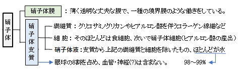
Its composition is similar to that of the cornea, but the vitreous contains very few cells. It is composed mostly of phagocytes, which remove unwanted cellular debris in the visual field, and hyalocytes, which turn over the hyaluronan.
The vitreous humour contains no blood vessels, and 98–99% of its volume is water (as opposed to only 75% in the cornea). In addition to water, the vitreous consists of salts, sugars, vitrosin (a type of collagen), a network of collagen type II fibrils with glycosaminoglycan, hyaluronan, opticin, and a wide array of proteins. Despite having little solid matter, the fluid is substantial enough to fill the eye and give it its spherical shape. This contrasts with the aqueous humour, which is more fluid, and the lens, on the other hand, which is elastic in nature and is tightly packed with cells. The vitreous humour has a viscosity two to four times that of water, giving it a gelatinous consistency. It has a refractive index of 1.336.[10]
【 語 句 】
・cornea:角膜 ・phagocyte:食細胞 ・debris:破片 ・hyalocyte:硝子体細胞 ・hyaluronan:ヒアルロン酸 ・spherical:球形の ・aqueous humour:房水 ・viscosity:粘度 ・consistency:濃度 ・refractive index:屈折率
【Development】
The vitreous fluid is not present at birth (the eye being filled with only the gel-like vitreous body), but found after age 4-5, and increases in size thereafter.[1]
Produced by cells in the non-pigmented portion of the ciliary body, the vitreous humour is derived from embryonic mesenchyme cells, which degenerate after birth.[1]
The nature and composition of the vitreous humour changes over the course of life. In adolescence, the vitreous cortex becomes more dense and vitreous tracts develop; and in adulthood, the tracts become better defined and sinuous. Central vitreous liquefies, fibrillar degeneration occurs, and the tracts break up (syneresis).[citation needed]
Coarse strands develop with ageing. The gel volume decreases with age, and the liquid volume increases.[12] The cortex may disappear at sites, allowing liquid vitreous to extrude adjacently into the potential space between vitreous cortex and retina (vitreous detachment).
【 語 句 】
・: ・: ・: ・: ・: ・: ・: ・: ・: ・: ・: ・: ・: ・: ・: ・: ・: ・: ・: ・: ・: ・: ・: ・: ・: ・: ・: ・: ・: ・: ・: ・: ・: ・: ・: ・:
■ 写真やイラストを掲載しているサイト ■
・ イラストや写真を掲載しているサイト-Ⅰ
・ イラストや写真を掲載しているサイト-Ⅱ
・ イラストや写真を掲載しているサイト-Ⅲ
・ イラストや写真を掲載しているサイト-Ⅳ
・ イラストや写真を掲載しているサイト-Ⅴ