※ちょっとどの資料だかは定かではないのだが、資料によっては「硝子体動脈の跡は完全に消滅する」
という解説文があったのを記憶している。また、インターネットで「眼球」の画像検索をしてみると、この
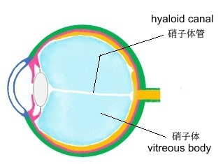
硝子体管を描いているイラストと全く描いていないイラストに分かれる。
以下は「世界大百科事典」の「硝子体」の解説文の一部となる。
「胎生第6週~7ヵ月までの間,ここには硝子体動脈があって,水晶体などに血液を供給するが,8ヵ月までには大部分消失する。 」
以下は「Wikipedia」の解説文となる。
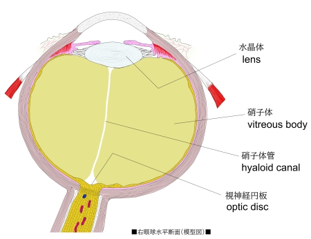
Hyaloid canal (Cloquet's canal and Stilling's canal) is a small transparent canal running through the vitreous body from the optic nerve disc (at the punctum caecum) to the lens. It is formed by an invagination of the hyaloid membrane, which encloses the vitreous body.
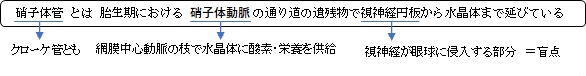
In the fetus, the hyaloid canal contains a prolongation of the central artery of the retina, the hyaloid artery, which supplies blood to the developing lens. Once the lens is fully developed the hyaloid artery retracts and the hyaloid canal contains lymph. The hyaloid canal appears to have no function in the adult eye, though its remnant structure can be seen.
Contrary to initial belief, the hyaloid canal does not facilitate changes in the volume of the lens. The lens volume changes by less than 1% over its range of accommodation. Furthermore, lymph, being liquid, is incompressible, so even if the volume of the lens did change, the hyaloid canal could not compensate for it.
【 語 句 】
・transparent:透明な ・vitreous body:硝子体 ・optic erve disc:視神経円板 ・punctum caecum:盲点 ・invagination:嵌入 ・hyaloid membrane:硝子体膜 ・enclose:包み込む ・fetus:胎児 ・prolongation:延長 ・central artery of the retina:網膜中心動脈 ・hyaloid artery:硝子体動脈 ・retract:縮む ・remnant:異物 ・facilitate:促進する ・range of accommodation:調節幅 ・incompressible:圧縮できない ・compensate:補う
■ 写真やイラストを掲載しているサイト ■
・ イラストや写真を掲載しているサイト-Ⅰ
・ イラストや写真を掲載しているサイト-Ⅱ
・ イラストや写真を掲載しているサイト-Ⅲ
・ イラストや写真を掲載しているサイト-Ⅳ