仙骨神経とは

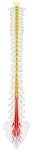
以下は脊髄神経を簡単に表した図となる。
脊髄神経
spinal nerves |
1 |
頚神経 cervical nerves |
8 対 |
第1頸神経 ~ 第8頸神経( C 1~ C8 ) |
2 |
胸神経 thoracic nerves |
12 対 |
第1胸神経 ~ 第12胸神経( T1 ~ T12 ) |
3 |
腰神経 lumbal nerves |
5 対 |
第1腰神経 ~ 第5腰神経( L1 ~ L5 ) |
4 |
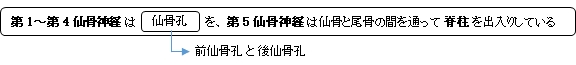
仙骨神経 sacral nerves |
5 対 |
第1仙骨神経 ~ 第5仙骨神経( S1 ~ S5 ) |
5 |
尾骨神経 coccygeal nerve |
1 対 |
尾骨神経( CO ) |
以下は「 Wikipedia 」の解説文の一部になる。
「 The roots of these nerves begin inside the vertebral column at the level of the L1 vertebra, where the cauda equina(馬尾)begins, and then descend into the sacrum. 」
※「 these nerves = sacral nerves 」
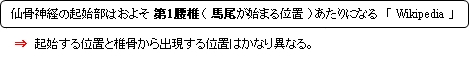
⇒ 仙骨神経の起始部が第1腰椎あたりであることが分かるイラストを掲載しているサイト

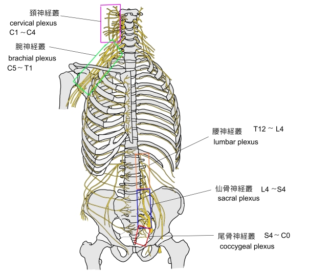
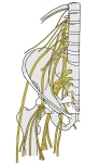
以下は脊髄神経が形成する神経叢を簡単に表した図になる。

腰仙骨神経叢(模型図) |
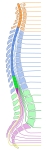
脊髄神経・起始部
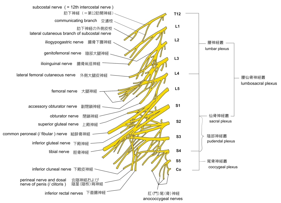
模型図 - 2 |
腰神経~尾骨神経 |
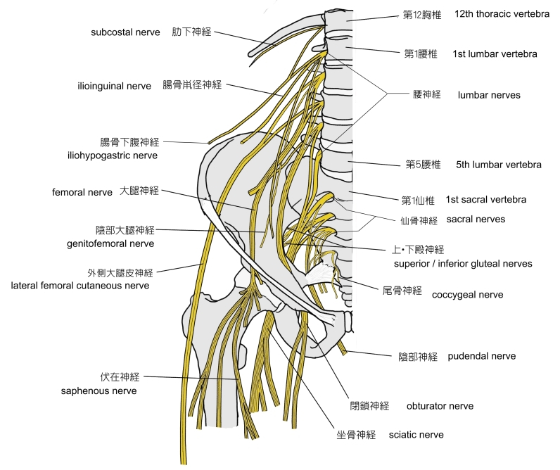
脊髄神経・起始部
模型図 - 3 |
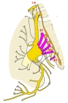
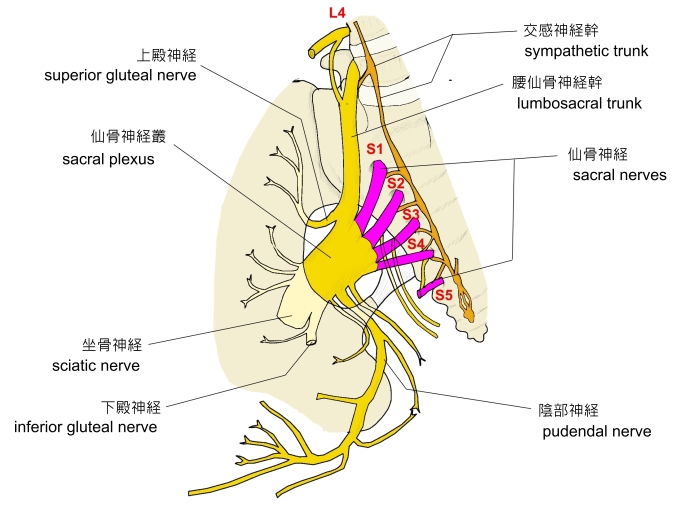
仙骨神経・仙骨神経叢
|

「 船戸和弥のホームページ 」では以下のように解説している。
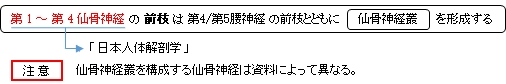
「 仙骨神経は5対あって、それぞれ、前枝と後枝とに分かれる。 前枝は前仙骨孔を通って出る。S1・S2・S3・S4・はL4・L5とともに仙骨神経叢をつくる。 後枝は後仙骨孔を通って出る。第1~3仙骨神経(S1・S2・S3)の後枝の外側枝は中殿皮神経として殿部の中部の皮膚に分布する。(解剖学講義)
・ 内側枝 :(仙骨神経の内側枝は脊髄神経の最終枝で多裂筋および仙骨と尾骨上の皮膚へいたる。)
・ 外側枝 : 仙骨神経の外側枝または尾骨神経の外側枝は尾骨上の皮膚へいたる知覚枝。S1~S3よりの後枝。)
・ 後皮枝 ; 後皮神経:仙骨神経の後皮枝はS1~S3より起る。大臀筋を貫き、上内側臀部の皮膚へ分布する。)
」
また、以下は「 Wikipedia 」の解説文となる。
「 The sacral nerves are the five pairs of spinal nerves which exit the sacrum at the lower end of the vertebral column. The roots of these nerves begin inside the vertebral column at the level of the L1 vertebra, where the cauda equina begins, and then descend into the sacrum.[2][3]
There are five paired sacral nerves, half of them arising through the sacrum on the left side and the other half on the right side. Each nerve emerges in two divisions: one division through the anterior sacral foramina and the other division through the posterior sacral foramina.[4]
The nerves divide into branches and the branches from different nerves join with one another, some of them also joining with lumbar or coccygeal nerve branches. These anastomoses of nerves form the sacral plexus and the lumbosacral plexus. The branches of these plexus give rise to nerves that supply much of the hip, thigh, leg and foot.[5][6]
The sacral nerves have both afferent and efferent fibers, thus they are responsible for part of the sensory perception and the movements of the lower extremities of the human body. From the S2, S3 and S4 arise the pudendal nerve and parasympathetic fibers whose electrical potential supply the descending colon and rectum, urinary bladder and genital organs. These pathways have both afferent and efferent fibers and, this way, they are responsible for conduction of sensory information from these pelvic organs to the central nervous system (CNS) and motor impulses from the CNS to the pelvis that control the movements of these pelvic organs.[7]」
【 語 句 】
・ cauda equina : 馬尾 ・ anterior sacral foramina : 前仙骨孔 ・ anastomose : 吻合 ・ lumbosacral plexus : 腰仙骨神経叢 ・ afferent : 求心性の ・ efferent : 遠心性の ・ sensory perception : 知覚認知 ・ lower extremities : 下肢 ・ pudendalnerve : 陰部神経 ・ parasympathetic fiber : 副交感神経線維 ・ electrical potential : 電位 ・ descending colon : 下行結腸 ・ rectum : 直腸 ・ urinary bladder : 膀胱 ・ genital organ : 生殖器 ・ conduction : 伝達
【 イラスト掲載サイト 】
・イラストや写真を掲載しているサイト-Ⅰ
・イラストや写真を掲載しているサイト-Ⅱ
・イラストや写真を掲載しているサイト-Ⅲ
・イラストや写真を掲載しているサイト-Ⅳ

|