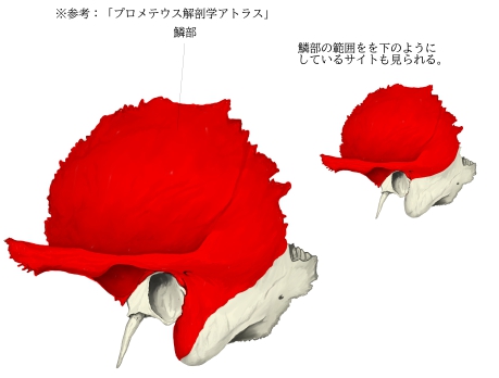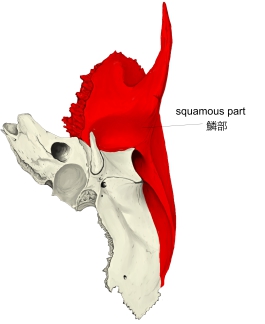
・「The squamous part of temporal bone is scale-like, thin, and translucent.」(Wikipedia)
scale:ウロコ translucent:半透明の
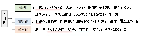
1 |
外(側)面 |
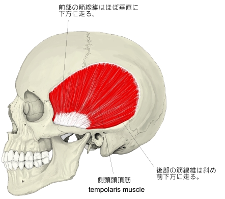
=側頭面
・滑らかで凸状となる。
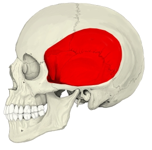
・側頭筋の起始部となっている。
・側頭窩の一部となる。
・後方に中側頭動脈溝が縦に走っている。
・後部下部に上下2本の側頭線が確認でき、上側頭線は側頭筋膜の付着部となっている。
・外耳孔の上部からほぼ水平に前方に頬骨突起が伸び、頬骨の側頭突起と接合して頬骨弓を形成する。前下部より咬筋が起始する。
・外耳道上壁の入口に接して尖った小突起を明瞭に認めるとき、これを道上棘という。
|
2 |
内(側)面 |
=大脳面
「わずかに陥凹して脳隆起、指圧痕、動・静脈溝を示し、ことに中硬膜静脈溝は明瞭である。大脳面と錐体部との間には錐体鱗裂があって、錐体部と鱗部との癒合を示すもので、加齢とともに次第に不明瞭となる。」(日本人体解剖学)
|
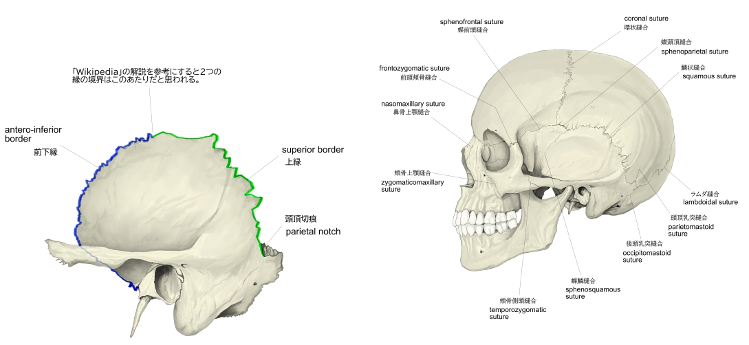
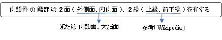
以下の縁の解説は「Wikipedia」を参考にしたものとなる。
1 |
上縁 |
・薄い。
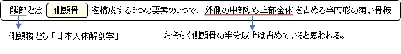
・前部:蝶形骨の大翼との縫合 ⇒蝶鱗縫合
・頭頂切痕を形成し、頭頂骨の乳突角が嵌入する。 |
|
2 |
前下縁 |
・厚く、鋸歯状となる。
・頭頂骨との縫合 ⇒鱗状縫合 |
以下は「Wikipedia」の解説文となる。
The squamous part of temporal bone, or temporal squama, forms the front and upper part of the temporal bone, and is scale-like, thin, and translucent.
【Surfaces】
Its outer surface is smooth and convex; it affords attachment to the temporal muscle, and forms part of the temporal fossa; on its hinder part is a vertical groove for the middle temporal artery. A curved line, the temporal line, or supramastoid crest, runs backward and upward across its posterior part; it serves for the attachment of the temporal fascia, and limits the origin of the temporalis muscle. The boundary between the squamous part and the mastoid portion of the bone, as indicated by traces of the original suture, lies about 1 cm. below this line.
【 語 句 】
・scale:鱗 ・translucent:半透明の ・convex:凸状の ・temporal muscle:側頭筋 ・temporal fossa:側頭窩 ・hinder:後方の ・groove:溝 ・middle temporal artery:中側頭動脈 ・fascia:筋膜 ・mastoid portion:乳突部 ・suture:縫合
Projecting from the lower part of the squamous part is a long, arched process, the zygomatic process. This process is at first directed lateralward, its two surfaces looking upward and downward; it then appears as if twisted inward upon itself, and runs forward, its surfaces now looking medialward and lateralward. The superior border is long, thin, and sharp, and serves for the attachment of the temporal fascia; the inferior, short, thick, and arched, has attached to it some fibers of the masseter. The lateral surface is convex and subcutaneous; the medial is concave, and affords attachment to the masseter. The anterior end is deeply serrated and articulates with the zygomatic bone. The posterior end is connected to the squamous part by two roots, the anterior and posterior roots. The posterior root, a prolongation of the upper border, is strongly marked; it runs backward above the external auditory meatus, and is continuous with the temporal line. The anterior root, continuous with the lower border, is short but broad and strong; it is directed medialward and ends in a rounded eminence, the articular tubercle (eminentia articularis).
【 語 句 】
・projecting:出っ張り ・zygomatic process:頬骨突起 ・masseter:咬筋 ・subcutaneous:皮下の ・concave:凹状の ・serrated:鋸歯状の articulate with~:~と接合する ・prolongation:延長 ・external auditory meatus:外耳道 ・eminence:高くなった部分 ・articular tubercle:関節結節
This tubercle forms the front boundary of the mandibular fossa, and in the fresh state is covered with cartilage. In front of the articular tubercle is a small triangular area which assists in forming the infratemporal fossa; this area is separated from the outer surface of the squamous part by a ridge which is continuous behind with the anterior root of the zygomatic process, and in front, in the articulated skull, with the infratemporal crest on the great wing of the sphenoid. Between the posterior wall of the external acoustic meatus and the posterior root of the zygomatic process is the area called the suprameatal triangle (Macewen), or mastoid fossa, through which an instrument may be pushed into the tympanic antrum.
【 語 句 】
・mandibular fossa:下顎窩 ・cartilage:軟骨 ・infratemporal fossa:側頭下窩 ・ridge:隆起(線) ・infratemporal crest:側頭下稜 ・great wing of the sphenoid:蝶形骨の大翼 ・suprameatal triangle:オトガイ上三角 ・mastoid fossa:乳突窩 ・instrument:器具? ・tympanic antrum:鼓室前庭
At the junction of the anterior root with the zygomatic process is a projection for the attachment of the temporomandibular ligament; and behind the anterior root is an oval depression, forming part of the mandibular fossa, for the reception of the condyle of the mandible. The mandibular fossa (glenoid fossa) is bounded, in front, by the articular tubercle; behind, by the tympanic part of the bone, which separates it from the external acoustic meatus; it is divided into two parts by a narrow slit, the petrotympanic fissure (Glaserian fissure). The anterior part, formed by the squamous part, is smooth, covered in the fresh state with cartilage, and articulates with the condyle of the mandible. Behind this part of the fossa is a small conical eminence; this is the representative of a prominent tubercle which, in some mammals, descends behind the condyle of the mandible, and prevents its backward displacement. The posterior part of the mandibular fossa, formed by the tympanic part of the bone, is non-articular, and sometimes lodges a portion of the parotid gland.
【 語 句 】
・temporomandibular ligament:顎関節靭帯 ・oval:楕円形の ・condyle:顆状突起 ・mandible:下顎骨 ・tympanic part:鼓室部 ・petrotympanic fissure:錐体鼓室裂 ・conical:円錐の ・displacement:置換 ・parotid gland:耳下腺
The petrotympanic fissure leads into the middle ear or tympanic cavity; it lodges the anterior process of the malleus, and transmits the tympanic branch of the internal maxillary artery. The chorda tympani nerve passes through a canal (canal of Huguier), separated from the anterior edge of the petrotympanic fissure by a thin scale of bone and situated on the lateral side of the auditory tube, in the retiring angle between the squamous part and the petrous portion of the temporal bone.
The internal surface of the squamous part is concave; it presents depressions corresponding to the convolutions of the temporal lobe of the brain, and grooves for the branches of the middle meningeal vessels.
【 語 句 】
・tympanic cavity:鼓室 ・malleus:ツチ骨 ・maxillary artery:顎動脈 ・chorda tympani nerve:鼓索神経 ・auditory tube:耳管 ・convolution:回旋 ・temporal lobe:側頭葉 ・middle meningeal vessels:中硬膜動・静脈
【Borders】
The superior border is thin, and bevelled at the expense of the internal table, so as to overlap the squamous border of the parietal bone, forming with it the squamosal suture. Posteriorly, the superior border forms an angle, the parietal notch, with the mastoid portion of the bone.
The antero-inferior border is thick, serrated, and bevelled at the expense of the inner table above and of the outer below, for articulation with the great wing of the sphenoid.
【 語 句 】
・bevel:斜角をつける ・at the expense of~:~を犠牲にして ・parietal bone:頭頂骨 ・parietal notch:頭頂切痕 ・mastoid portion:乳突部
■ 写真やイラストを掲載しているサイト ■
・ イラストや写真を掲載しているサイト-Ⅰ
・ イラストや写真を掲載しているサイト-Ⅱ
・ イラストや写真を掲載しているサイト-Ⅲ
・ イラストや写真を掲載しているサイト-Ⅳ
・ イラストや写真を掲載しているサイト-Ⅴ