そして、「船戸和弥のホームページ」や「日本人体解剖学 (上巻) 」でははっきりとは言及していないが、以下のように考えてよいと思われる。 」でははっきりとは言及していないが、以下のように考えてよいと思われる。
■ 付着するもの ■
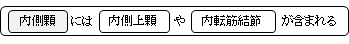
以下の一覧には内側上顆および内転筋結節に付着するものも挙げているので重複する。
筋 肉
|
1 |
|
|
peroneal muscle |
2 |
|
|
adductor magnus muscle |
靭 帯
|
1 |
|
|
medial collateral ligament |
2 |
|
内側顆 |
posterior cruciate ligament |
「船戸和弥のホームページ」には以下のような解説が見られる。
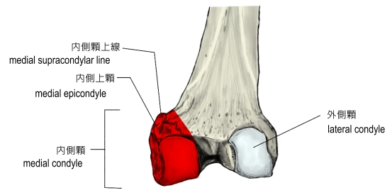
「大腿骨体の下部(遠位部)はことに著しく厚く大きく、その下端は左右の肥厚した内側顆および外側顆となる。内側顆は狭く長く、凸面の張り出しが強い。」
それに対して以下は「日本人体解剖学 (上巻) 」の解説文となる。
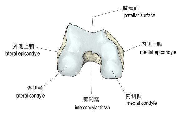
「(大腿骨の遠位端は)内側顆および外側顆という著しく肥大した部分で、前面には、膝蓋骨の後面と接する滑沢な膝蓋面がある。後面では、内側顆および外側顆の間は深く陥没し、これを顆間窩といい、上方は窩間線により膝窩平面から区別される。」
また、以下が「Wikipedia」の解説文となる。
「The medial condyle is one of the two projections on the lower extremity of femur, the other being the lateral condyle.
The medial condyle is larger than the lateral (outer) condyle due to more weight bearing caused by the centre of mass being medial to the knee. On the posterior surface of the condyle the linea aspera (a ridge running down the posterior shaft of the femur) turns into the medial supracondylar ridge. The outermost protrusion on the medial surface of the medial condyle is referred to as the "medial epicondyle" and can be palpated by running fingers medially from the patella with the knee in flexion.」
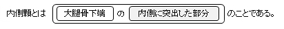
・内側顆は外側顆よりも大きい。
・後面で粗線が内側顆上線に移行している。
・最外側部の突出部を「内側上顆」という。
【参考になるサイト】
・イラストや写真を掲載しているサイト-Ⅰ(内側顆を色分け)
・イラストや写真を掲載しているサイト-Ⅱ(内側顆を色分けしているが、内側上顆および内転筋結節は含まず)
・イラストや写真を掲載しているサイト-Ⅲ
・イラストや写真を掲載しているサイト-Ⅳ

|