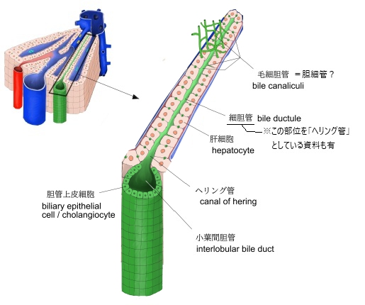

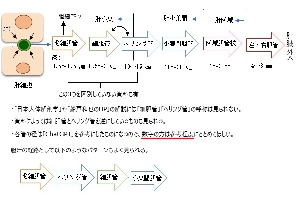
・毛細胆管は肝小葉の中を縦・横網目状に走っている。
・毛細胆管の径はChatGPT調べで「0.5~1.5㎛」ほどとなる。
・毛細胆管=胆細管、「ChatGPT」にこの毛細血管について尋ねると、以下のような回答である。
「毛細胆管」という言葉は日本語では時折使われますが、正確にはこれは「胆細管(bile canaliculus)」を指していると考えられます。
・「日本人体解剖学」の解説ではその使用が確認できる。ただ、「船戸和也のHP」にはこの呼称は見当たらない。
以下は「 日本人体解剖学 」の解説文となる。
毛細胆管は、特別な壁をもたない肝細胞間の隙間であり、中心静脈に近い端から始まり、肝細胞索の間を走って肝小葉の周辺に達し、介在部に続く。
介在部は約25㎛の短い管で、単層の立方あるいは扁平な細胞からでき、この管は毛細胆管に比べて著しく太いので、また、膨大部とも呼ばれる。2~3個の介在部が集まり、肝小葉外に出て小葉間胆管に移行する。小葉間結合組織およびグリッソン被膜内にはリンパ管も見られる。毛細胆管は、一般の腺腔にあたるものであるが、他の腺と異なって、ただ2個の肝細胞により囲まれている。
また「Wikipedia」では以下のように解説している。
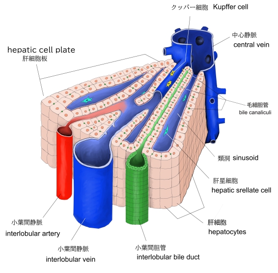
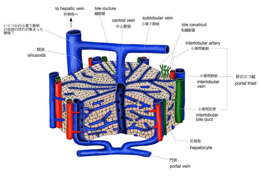
A bile canaliculus (pl.: bile canaliculi; also called bile capillaries) is a thin tube that collects bile secreted by hepatocytes. The bile canaliculi empty into a series of progressively larger bile ductules and ducts, which eventually become common hepatic duct. The bile canaliculi empty directly into the canals of Hering.
Hepatocytes are polyhedral in shape, therefore having no set shape or design, although they are made of cuboidal epithelial cells. They have surfaces facing the sinusoids (called sinusoidal faces) and surfaces which contact other hepatocytes (called lateral faces). Bile canaliculi are formed by grooves on some of the lateral faces of these hepatocytes.
Microvilli are present in the canaliculi.
【 語 句 】
・bile:胆汁 ・secrete:分泌する ・hepatocytes:肝細胞 ・bile ductules:細胆管 ・bile duct:胆管 ・common hepatic duct:総胆管 ・canals of Hering:ヘリング管 ・polyhedral:多面体の ・epithelial cells:上皮細胞 ・sinusoid:類洞 ・groove:溝 ・Microvilli:微絨毛
■ 写真やイラストを掲載しているサイト ■
・ イラストや写真を掲載しているサイト-Ⅰ