・「肝臓の実質は小葉間結合組織(グリソン鞘)によって多数の肝小葉に分けられる」(日本人体解剖学)
・「各小葉間の境界は、ルーペを使ってもそれほど明瞭ではない。」(船戸和也のHP)
理由:人間の肝小葉のグリソン鞘の発達が未熟なため。ブタなどは非常に明瞭の境界を示す。
⇒人間とブタの肝小葉の違いが分かる組織学的写真を掲載しているサイト

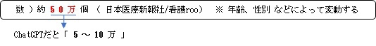
肝臓は約2500億から3000億の肝細胞で構成されているといわれている、この数を肝小葉の数で割ると
⇒ 「1つの肝小葉はおよそ50万ほどの肝細胞で構成されている」ことになる。
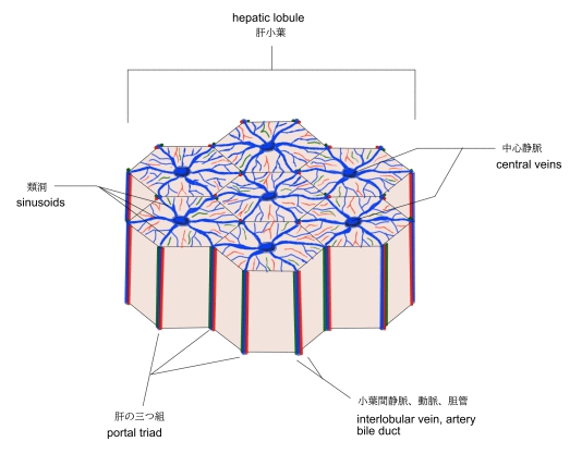
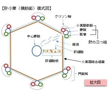
【構 造】
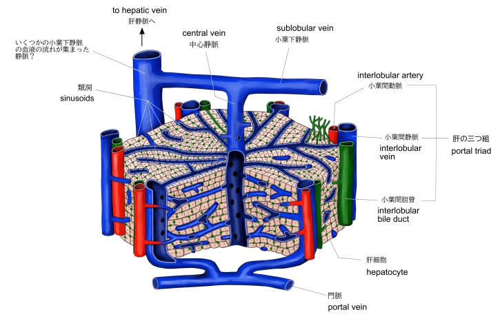
1. 角柱の各角にはグリソン鞘に包まれた小葉間静脈・動脈・胆管が位置し、この部位を門脈域と呼ぶ。
2. 小葉間動脈・静脈からの血液は類洞を経由して中心静脈へ注いでいる。
胆汁は血液とは逆の流れとなり、肝細胞で産出されたものが小葉間胆管に向って流れている。
3. 肝細胞は中心静脈に向って列をなしこれを肝細胞板と呼ぶ。
【参考となるサイト】
「船戸和也のHP」では以下のように解説している。
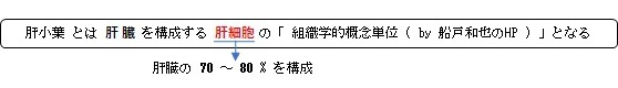
肝小葉は肝実質と血管は小さな肝臓の多角形の組織学的概念的単位で、肝静脈の1つの終末枝である中心静脈の周囲に存在する肝細胞集団からなり、周辺部には門脈枝、肝動脈、胆管がある。これら肝構造単位はどの導通路を構造単位の中心と考えるかによって、肝小葉であったり門脈小葉であったりする。
強靱な肝臓被膜から肝臓内へ、海綿状の繊細な結合組織が入っていく。これを足場としてこの中には、血管と胆管が走っており、結合組織の網目の中に肝上皮が横たわっている。肝臓の表面には数mm大の多角形の肝小葉がみえる。
肝小葉は多角形の表面をもつ(西洋)ナシの形をしており、大きさは断面で1~1.5mm、長さは約2mmである。これらの血管は肝小葉まわりで血管網を作るために枝を出す。この血管網から小葉へ枝が入っていき、これらの枝は門脈の血液を洞様血管へ導きいれる(小葉の周辺部は中心部より酸素の供給がよい)。基底部の(「ナシの柄」に相当する)からは中心静脈が出ていく。しかし、各小葉間の境界は、ルーペを使ってもそれほど明瞭ではない。これは小葉間を埋める結合組織すなわちグリソン鞘が人間ではあまり発達していないためである。ブタでは非常に明瞭。)
以下は「Wikipedia」の解説文となる。
The lobules of liver, or hepatic lobules, are small divisions of the liver defined at the microscopic (histological) scale. The hepatic lobule is a building block of the liver tissue, consisting of a portal triad, hepatocytes arranged in linear cords between a capillary network, and a central vein.
Lobules are different from the lobes of the liver: they are the smaller divisions of the lobes. The two-dimensional microarchitecture of the liver can be viewed from different perspectives:[1]
Name |
Shape |
Model |
classical lobule[2] |
hexagonal; divided into concentric centrilobular, midzonal, periportal parts |
anatomical |
portal lobule[3] |
triangular; centered on a portal triad |
bile secretion |
acinus [4] |
elliptical or diamond-shaped; divided into zone I (periportal), zone II (transition zone), and zone III (pericentral) |
blood flow and metabolic |
The term "hepatic lobule", without qualification, typically refers to the classical lobule.
The hepatic lobule can be described in terms of metabolic "zones", describing the hepatic acinus (terminal acinus). Each zone is centered on the line connecting two portal triads and extends outwards to the two adjacent central veins. The periportal zone I is nearest to the entering vascular supply and receives the most oxygenated blood, making it least sensitive to ischemic injury while making it very susceptible to viral hepatitis. Conversely, the centrilobular zone III has the poorest oxygenation, and will be most affected during a time of ischemia.[5]
Portal triad[edit]
A portal triad (also known as portal canal, portal field[citation needed], portal area[citation needed], or portal tract[citation needed]) is a distinctive arrangement within lobules. It consists of the following five structures:[6]
The misnomer "portal triad" traditionally has included only the first three structures, and was named before lymphatic vessels were discovered in the structure.[7][8] It can refer both to the largest branch of each of these vessels running inside the hepatoduodenal ligament, and to the smaller branches of these vessels inside the liver.
In the smaller portal triads, the four vessels lie in a network of connective tissue and are surrounded on all sides by hepatocytes. The ring of hepatocytes abutting the connective tissue of the triad is called the periportal limiting plate.
Periportal space[edit]
The periportal space (Latin: spatium periportale), or periportal space of Mall,[9] is a space between the stroma of the portal canal and the outermost hepatocytes in the hepatic lobule, and is thought to be one of the sites where lymph originates in the liver.[10]
Fluid (residual blood plasma) that is not taken up by hepatocytes drains into the periportal space, and is taken up by the lymphatic vessels that accompany the other portal triad constituents.
Function
Zones differ by function:
Other zonal injury patterns include zone I deposition of hemosiderin in hemochromatosis and zone II necrosis in yellow fever.[11]
【 語 句 】
・: ・: ・: ・: ・: ・: ・: ・: ・: ・: ・: ・: ・: ・: ・: ・: ・: ・: ・: ・: ・: ・: ・: ・: ・: ・: ・: ・: ・: ・: ・: ・:
■ 写真やイラストを掲載しているサイト ■
・ イラストや写真を掲載しているサイト-Ⅰ
・ イラストや写真を掲載しているサイト-Ⅱ
・ イラストや写真を掲載しているサイト-Ⅲ
・ イラストや写真を掲載しているサイト-Ⅳ