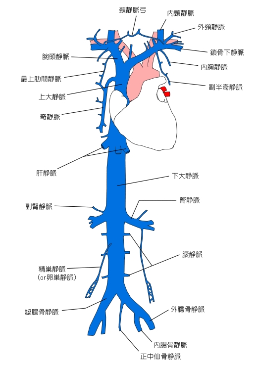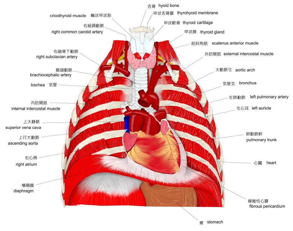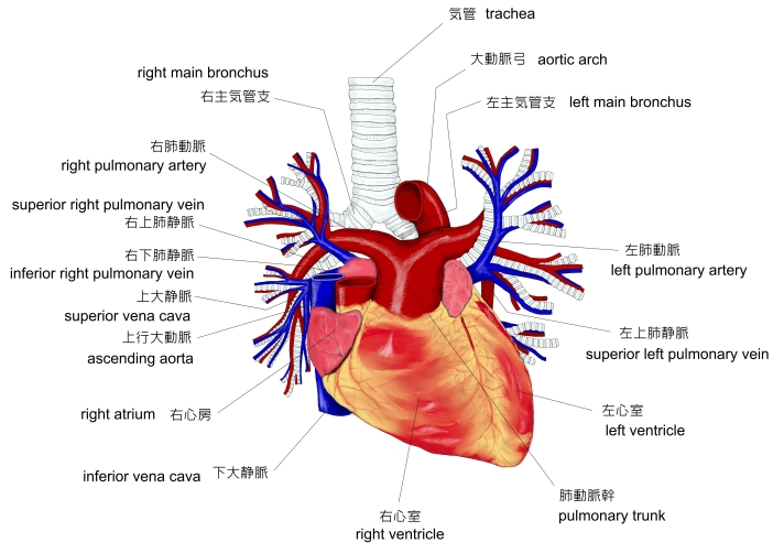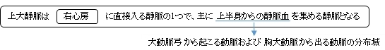
右心房 (right atrium) に直接入る静脈は上大静脈、下大静脈、そして冠状静脈洞の3つで、上大静脈は主に上半身からの静脈血 (venous blood) を集めて右心房に通じている。

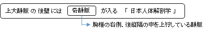
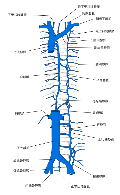
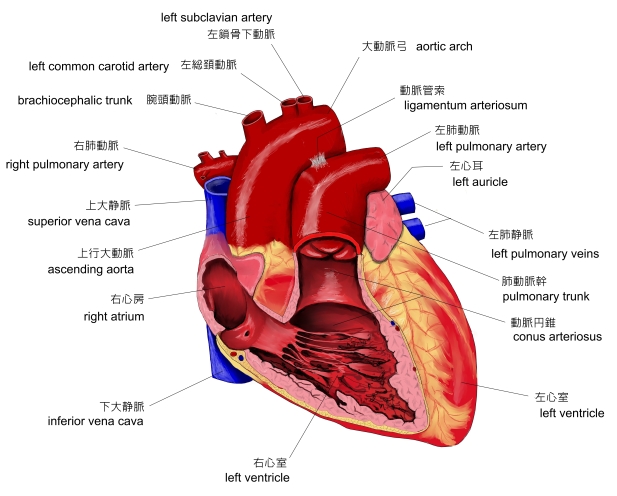
・ 奇静脈 (azygos vein) は第3胸椎(/第4胸椎)の高さで前方へ曲がり、右気管支 (right bronchus) の上を超えて上大静脈に入る。
以下は「 Rauber-Kopsch 解剖学 」の解説文の一部を箇条書きにしたものとなる。
・ 第2肋骨の高さでこの静脈は心膜によって不完全に包まれる.
・ 右側は胸膜の縦隔部で被われ,また右肺に策している.
・ 上大静脈は弁を持っていない.
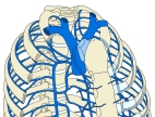

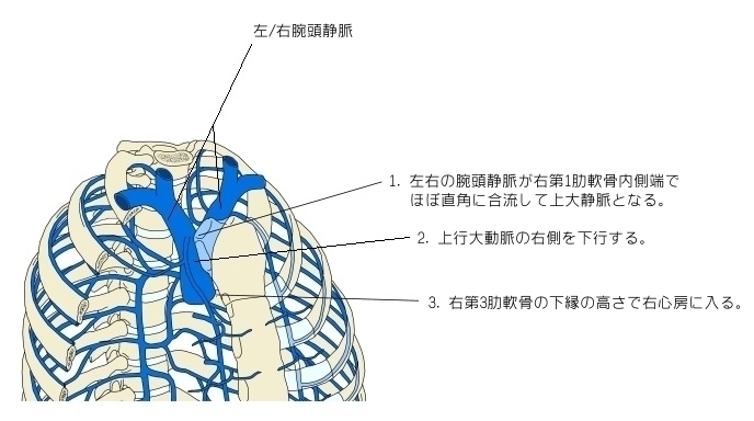
1 . 左右の腕頭静脈が右第1肋軟骨内側端で、ほぼ直角に合流して上大静脈となる。
2 . 上行大動脈の右側を下行する。
3 . 右第3肋軟骨の下縁の高さで右心房に入る。

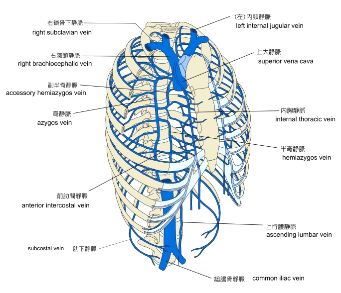
⇒ 肋軟骨と上大静脈の位置関係がよく分かるイラストを掲載しているサイト

以下は「 TeachMe Anatomy 」の解説文となる。
「 The superior vena cava is classified as a large vein, with a wide diameter of up to 2cm and a length of approximately 7cm.
It arises from the union of the left and right brachiocephalic veins, posterior to the first right costal cartilage. It descends vertically through the superior mediastinum, behind the intercostal spaces and to the right of the aorta and trachea.
At the level of the second costal cartilage, the SVC enters the middle mediastinum and becomes surrounded by the fibrous pericardium. It terminates by emptying into the superior aspect of the right atrium at the level of the third costal cartilage. 」
【 語 句 】
・ brachiocephalic veins : 腕頭静脈 ・ costal cartilage : 肋軟骨 ・ mediastinum : 縦隔 ・ intercostal spaces : 肋間隙 ・ aorta : 大動脈 ・ trachea : 気管 ・ fibrous pericardium : 線維性心膜 ・ right atrium : 右心房
【 イラスト掲載サイト 】
・ イラストや写真を掲載しているサイト-Ⅰ
・ イラストや写真を掲載しているサイト-Ⅱ
・ イラストや写真を掲載しているサイト-Ⅲ
・ イラストや写真を掲載しているサイト-Ⅳ
・ イラストや写真を掲載しているサイト-Ⅴ

|