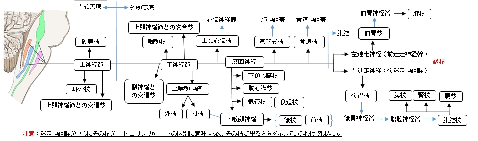
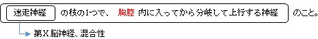
反回神経(迷走神経の枝)とは
走 行 )左右の反回神経は、その起始部の走行の仕方が少し異なる。
右反回神経 : 鎖骨下動脈 の下から後方に回る。
左反回神経 : 大動脈弓(動脈管索の外側)の下から後ろに回る。
それぞれ後方に回った後は
気管食道溝を上行し下咽頭収縮筋の下縁で下喉頭神経となって多数の枝に分岐する。
分 布 )筋 : 輪状甲状筋を除くすべての喉頭筋 ※輪状甲状筋は上喉頭神経の支配となる。
粘膜 : 喉頭下半の粘膜
吻 合 )上喉頭神経(上喉頭神経の内枝との交通枝)
枝 )下頚心臓枝、胸心臓枝、気管枝、食道枝、下喉頭神経(終枝)
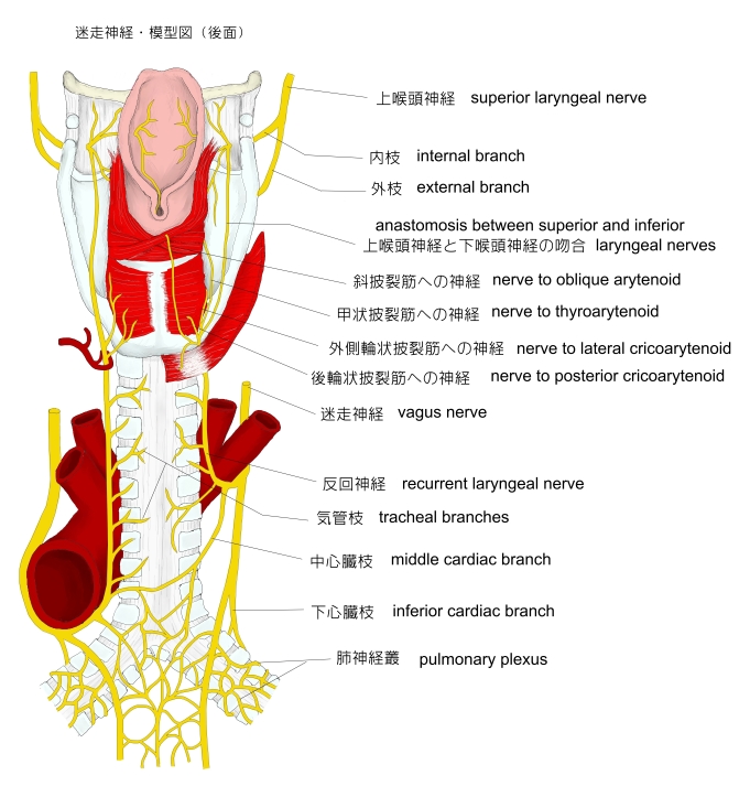
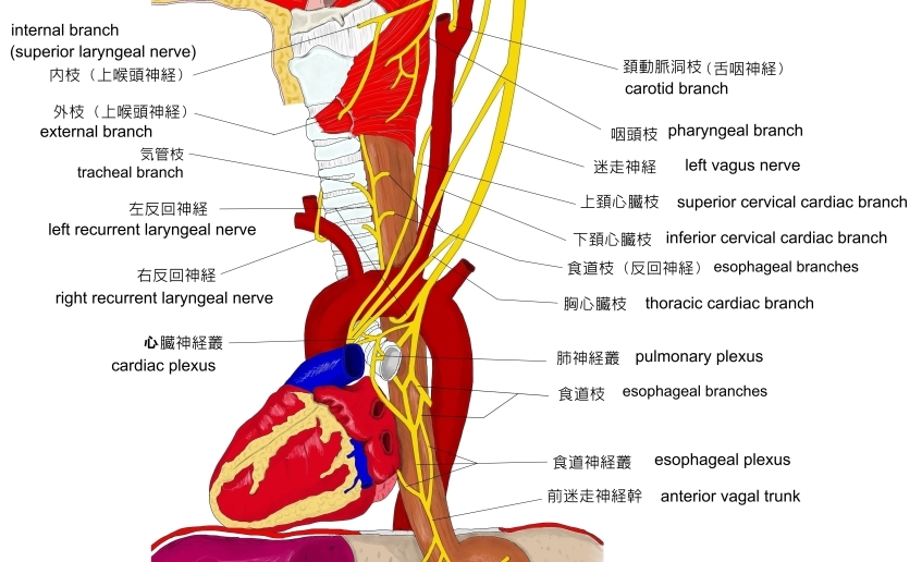
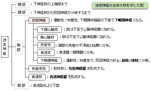
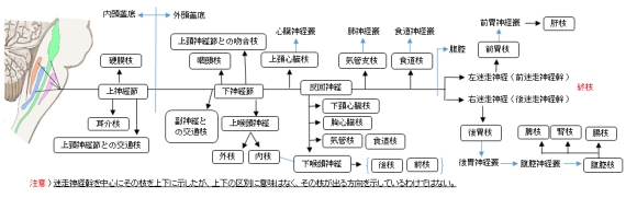
以下は迷走神経の4つの区分と胸部の構造を簡単に表した図になる。
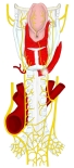
迷走神経・模型図
(頚部/胸部後面)
|
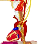
迷走神経模型図
(頚部)
|
|
|
【参考になるサイト】
・イラストや写真を掲載しているサイト-Ⅰ
・イラストや写真を掲載しているサイト-Ⅱ
・イラストや写真を掲載しているサイト-Ⅲ
・イラストや写真を掲載しているサイト-Ⅳ
|