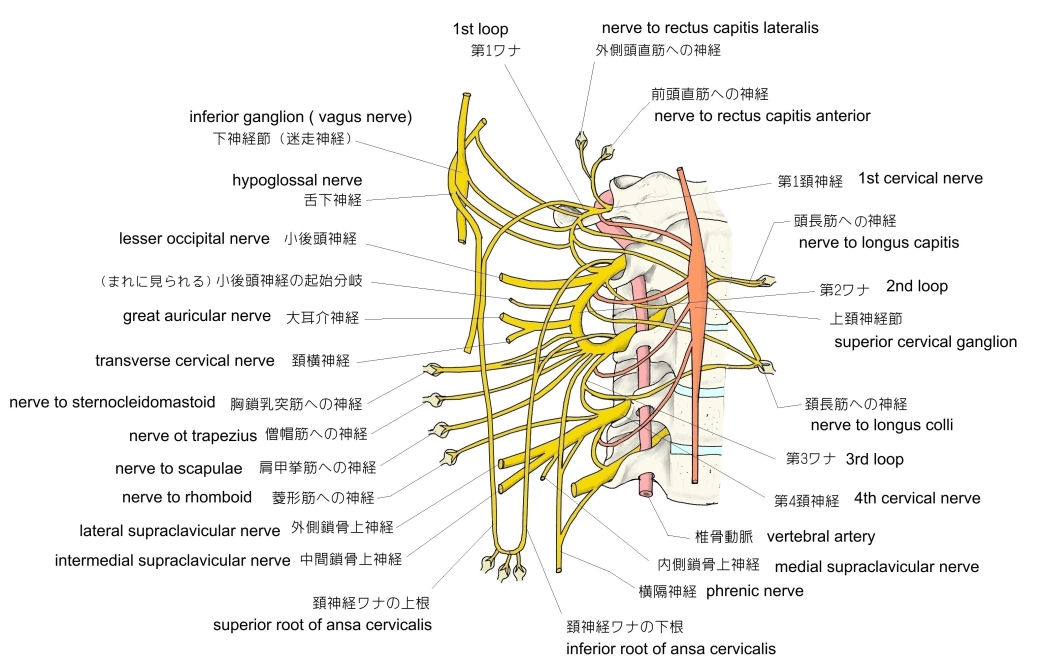
以下は頚神経叢の起始や枝を簡単に表した図になる。

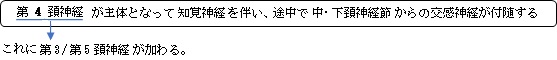
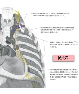
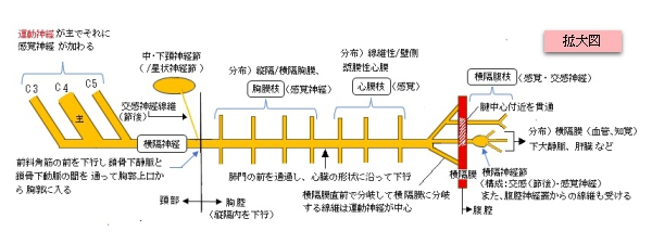
1. 第4頸神経(C4)を主とし、第3(C3)および第5(C5)頸神経からの枝が合流して起始する。
( 起始部では運動神経線維と知覚神経線維からなる脊髄神経となる。)
2. 前斜角筋の前面外側から内側縁に向かって斜めに下行する。
(頸部の下端(鎖骨のすぐ上付近)で、中・下頸神経節(あるいは星状神経節)からの交通枝を介して交感神経線維が加わる。)
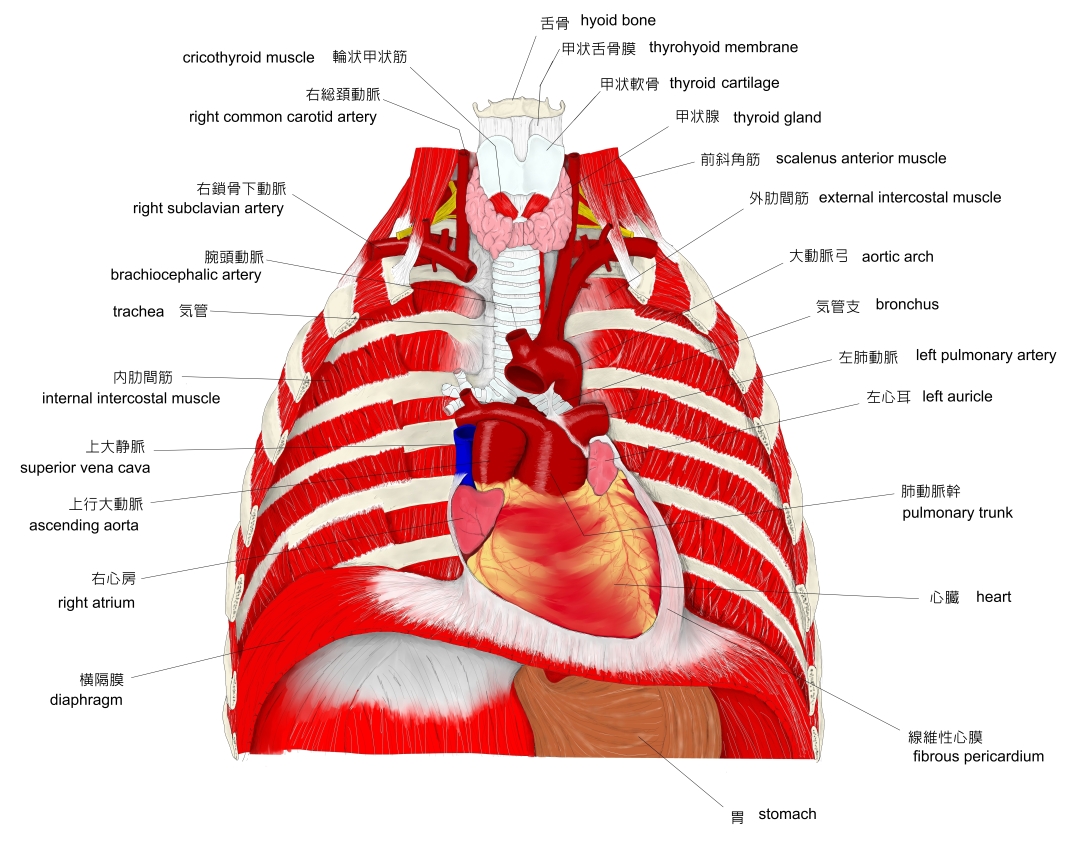
3. 胸郭上口にて、鎖骨下動脈の前面、かつ鎖骨下静脈の後面を通って胸腔内に入る。
(胸腔内からは(それ以前もその傾向はあるが…)前下方に走行する形になるが、左側は右側に比べ、心臓の膨らみに沿うため、前下方への傾斜が強い。)
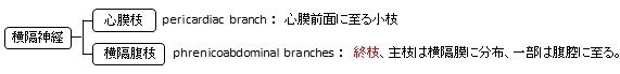
4. 縦隔において、縦隔胸膜と線維性心膜の間に挟まれるように、心膜横隔動脈・静脈を伴走させて下行する。
(下行中、多くの細かな枝(胸膜枝・心膜枝)を出し、胸膜、心膜、および横隔膜中央部の知覚を司る。)
5. 左右とも肺門の前方を通り、心臓の側面に達すると、心膜(線維性心膜および壁側漿膜)へ分布する心膜枝を分岐しながら心臓の形状に沿って下行する。
6. 横隔膜に達すると数本に分岐し、一部の終末枝は横隔膜の上面を放射線状に広がって筋腹に分布する。
7. 別の終末枝(横隔腹枝)は、大静脈孔(右側)を通過したり、あるいは直接筋部を貫通(左側)して横隔膜の下面(腹面)に回り、腹膜や周辺の靭帯、下大静脈、副腎付近の知覚を司る。
・走行がよく分かるイラスト掲載サイト-1

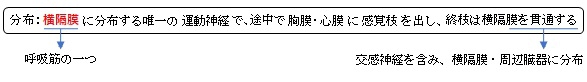
横隔神経は混合神経だが主は運動神経で、縦隔を下行し横隔膜に到達後、扇状に分岐して筋線維に分布。その走行の際に以下の枝(感覚神経、交感神経)を出す。

以下は「 Wikipedia 」の解説文となる。
「 The phrenic nerve is a nerve that originates in the neck (C3-C5) and passes down between the lung and heart to reach the diaphragm. It is important for breathing, as it passes motor information to the diaphragm and receives sensory information from it. There are two phrenic nerves, a left and a right one.
The phrenic nerve originates mainly from the 4th cervical nerve, but also receives contributions from the 5th and 3rd cervical nerves (C3-C5) in humans.[1] Thus, the phrenic nerve receives innervation from parts of both the cervical plexus and the brachial plexus of nerves.
The phrenic nerves contain motor, sensory, and sympathetic nerve fibers. These nerves provide the only motor supply to the diaphragm as well as sensation to the central tendon. In the thorax, each phrenic nerve supplies the mediastinal pleura and pericardium.
Structure
The phrenic nerve descends obliquely with the internal jugular vein across the anterior scalene, deep to the prevertebral layer of deep cervical fascia and the transverse cervical and suprascapular arteries. On the left, the phrenic nerve crosses anterior to the first part of the subclavian artery. On the right, it lies on the anterior scalene muscle and crosses anterior to the 2nd part of the subclavian artery. On both sides, the phrenic nerve runs posterior to the subclavian vein as it enters the thorax where it runs anterior to the root of the lung and between the fibrous pericardium and mediastinal face of the parietal pleura.[1]
Found in the middle mediastinum, both phrenic nerves run from C3, C4, and C5 along the anterior scalene muscle deep to the carotid sheath.
The pericardiacophrenic arteries and veins travel with their respective phrenic nerves.
The phrenic nerve can be marked by a line connecting these two points:
- 1st point can be labelled 3.5 cm at the level of the thyroid cartilage from the midsagittal plane.
- 2nd point is at the medial end of the clavicle.
Variation
The contribution of the 5th cervical nerve may stem from an accessory phrenic nerve. Phrenic nerve in its early course close to its origin, was giving a communicating branch to C5 root of brachial plexus. The phrenic nerve at the level of root of neck just before entering the thorax was placed in front of the subclavian vein. Usually it is placed posterior in between subclavian vein and artery.[2]
Most often it is a branch of the nerve to the subclavius and may contain numerous phrenic nerve fibers. If the accessory phrenic nerve is present, it lies lateral to the main nerve and descends posterior and occasionally inferior to the subclavian vein. The accessory phrenic nerve connects to the phrenic nerve in the thorax or the root of the neck.[1]
In canines the phrenic nerve arises from C5-C7 with occasional small contributions from C4.[3] In the cat, horse, ox, and small ruminant the phrenic nerve arises variably from C4-C7.
Function
Both of these nerves supply motor fibers to the diaphragm and sensory fibers to the fibrous pericardium, mediastinal pleura, and diaphragmatic peritoneum.
Some sources describe the right phrenic nerve as innervating the gallbladder, other sources make no such mention.[4]」
【 語 句 】
・: ・: ・: ・: ・: ・: ・: ・: ・: ・: ・: ・:
【 イラスト掲載サイト 】
・イラストや写真を掲載しているサイト-Ⅰ
・イラストや写真を掲載しているサイト-Ⅱ
・イラストや写真を掲載しているサイト-Ⅲ
・イラストや写真を掲載しているサイト-Ⅳ
・イラストや写真を掲載しているサイト-Ⅴ

|