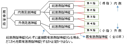
内側足底神経とは
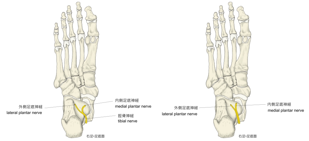
・上記では「足底で」と記したが「船戸和弥のホームページ 」では「 屈筋支帯の下で脛骨神経より起こり 」という解説が見られる。また、インターネットで画像検索をすると、足底を描いたスラストなどでは、明らかに足底で脛骨神経が内側足底神経と外側足底神経に分岐しているものを掲載しているサイトもあれば、足底部ではすでに2つに分岐しているイラストを掲載しているサイトも見受けられる。
「 船戸和弥のホームページ 」には以下のような解説文が見られる。
「 外側足底神経よりも大きく、手における正中神経と相同である。」
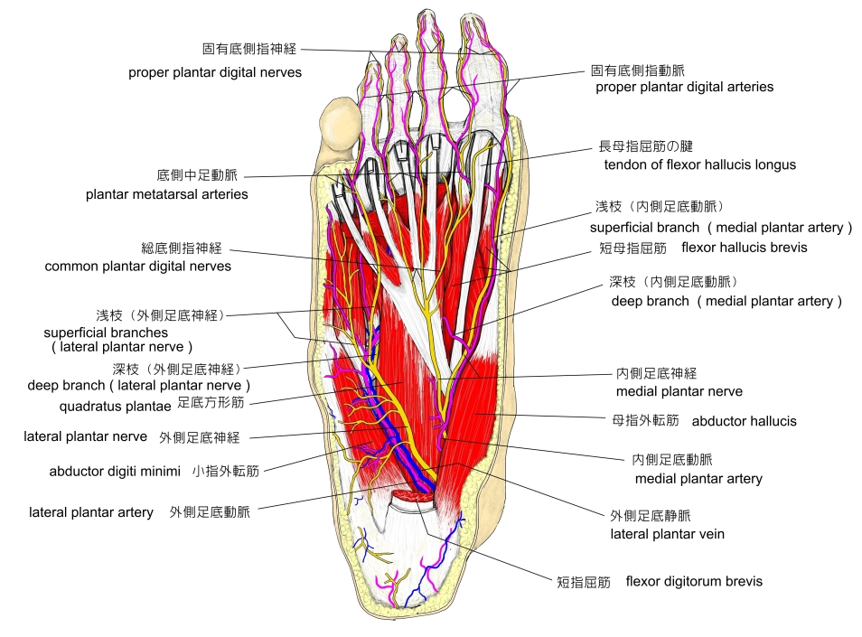
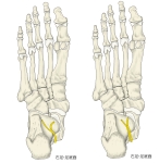
右足底の神経
|

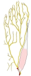
右足底の神経・筋肉
|
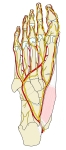
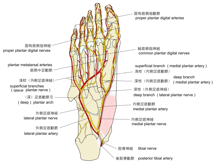
右足底の神経・動脈
|
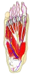
右足底の血管・神経・筋肉
|

以下は「 船戸和弥のホームページ 」を参考にしたものとなる。
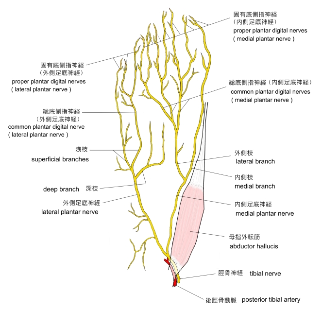
1 . 分岐後、母指外転筋の深部を内側足底動脈・静脈の外側に沿って走行
2 . 次に母指外転筋と短指屈筋の間を走行
3 . ほぼ足根中足関節の位置で 外側枝 と 内側枝 に分岐
・ 外側枝 : 3本の 総底側指神経 に分岐し、各総底側指神経はそれぞれ2本の固有底側指神経に分岐する。
・ 内側枝 : 母指の内側縁に向かう 固有底側指神経 となる。

以下は「 日本人体解剖学 (上巻) 」の解説文となる。 」の解説文となる。
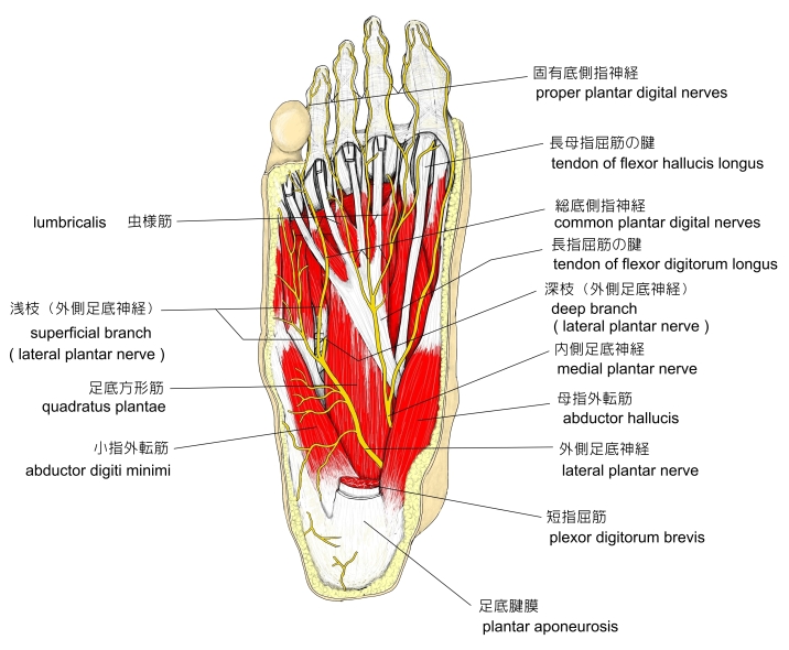
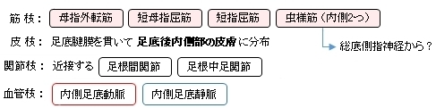
「 筋枝を母指球の筋(母指外転筋、短母指屈筋)、短指屈筋、内側の2つの虫様筋に与え、皮枝を足底内側の皮膚に与えたのち、内・外側の2枝に分かれる。」
ただ、「船戸和弥のホームページ 」では虫様筋への筋枝に関しては、分岐した総底側指神経が出している解説になっている。以下は、関節枝や血管枝に関しても解説している「 船戸和弥のホームページ 」を参考にして作成したものになる。
|
|
|
|
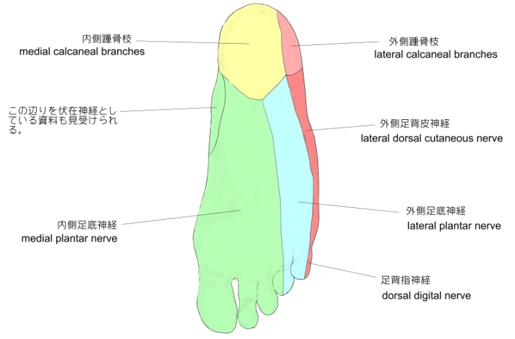
右足(底部)
|

内側足底神経は、およそ足根中足関節の位置で大きく内側枝と外側枝の2つに分岐する。内側枝は固有底側指神経として母指の内側縁の皮膚に至り、外側枝は3つの総底側指神経に分岐し、それぞれ総底側指神経が2つの固有底側指神経として分かれて母指の外側縁から第4指の内側縁の皮膚に至る。

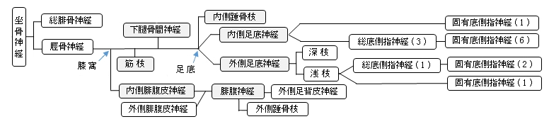
以下は脛骨神経全体の枝を表す簡単な図となる。


以下は「 Wikipedia 」の解説文となる。
「 The medial plantar nerve (internal plantar nerve) is the larger of the two terminal divisions of the tibial nerve (medial and lateral plantar nerve), which accompanies the medial plantar artery.
From its origin under the laciniate ligament it passes under cover of the abductor hallucis muscle , and, appearing between this muscle and the flexor digitorum brevis, gives off a proper digital plantar nerve and finally divides opposite the bases of the metatarsal bones into three common digital plantar nerves.
【 branches 】
The branches of the medial plantar nerve are: (1) cutaneous, (2) muscular, (3) articular, (4) a proper digital nerve to the medial side of the great toe, and (5) three common digital nerves.
【 cutaneous branches 】
The cutaneous branches pierce the plantar aponeurosis between the abductor hallucis and the flexor digitorum brevis and are distributed to the skin of the sole of the foot.
【 muscular branches 】
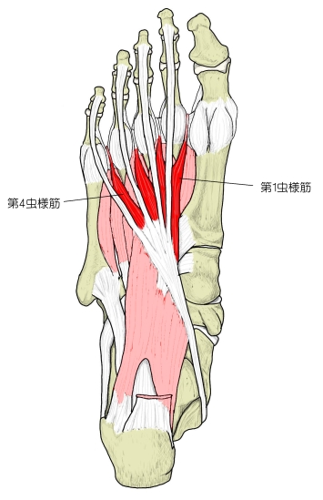
The muscular branches supply muscles on the medial side of the sole, including the abductor hallucis, the flexor digitorum brevis, the flexor hallucis brevis, and the first lumbrical; those for the abductor hallucis and flexor digitorum brevis arise from the trunk of the nerve near its origin and enter the deep surfaces of the muscles; the branch of the flexor hallucis brevis springs from the proper digital nerve to the medial side of the great toe, and that for the first lumbricalis from the first common digital nerve.
【 articular branches 】
The articular branches supply the articulations of the tarsus and metatarsus.
【 proper digital nerve of the great toe 】
The proper digital nerve of the great toe (nn. digitales plantares proprii; plantar digital branches) supplies the flexor hallucis brevis and the skin on the medial side of the great toe.
【 three common digital nerves 】
The three common digital nerves (nn. digitales plantares communes) pass between the divisions of the plantar aponeurosis, and each splits into two proper digital nerves—those of the first common digital nerve supply the adjacent sides of the great and second toes; those of the second, the adjacent sides of the second and third toes; and those of the third, the adjacent sides of the third and fourth toes.
The third common digital nerve receives a communicating branch from the lateral plantar nerve; the first gives a twig to the first lumbricals.
Each proper digital nerve gives off cutaneous and articular filaments; and opposite the last phalanx(指骨) sends upward a dorsal branch, which supplies the structures around the nail, the continuation of the nerve being distributed to the ball of the toe.
It will be observed that these digital nerves are similar in their distribution to those of the median nerve in the hand. 」
【 語 句 】
・ abductor hallucis muscle : 母指外転筋 ・ metatarsal bones : 中足骨 ・ tarsus : 足根骨 ・ metatarsus : 中足骨 ・ plantar aponeurosis : 足底腱膜 ・ adjacent : 隣接した ・ phalanx : 指骨
また、「 船戸和弥のホームページ 」では以下のように解説している。
「 内側足底神経は、外側足底神経よりも大きく、手における正中神経と相同である(図8)。この神経は、屈筋支帯の下で脛骨神経より起こり、始めは母趾外転筋の深部を内側足底動静脈の外側に沿って走り、ついで母趾外転筋と短趾屈筋の間を進み、最終的には短母趾屈筋と短趾屈筋の間を進み、最終的には短母趾屈筋と短趾屈筋との間を走行する。ほぼ足根中足関節のⅠで、内側足底神経は母趾内側皮膚へ行く固有底側趾神経と3本の総底側趾神経に分岐して終わる。
この分岐より以前に、内側足底神経は、母趾外転筋、短趾屈筋および短母趾屈筋への筋枝;足底腱膜を貫き足底後内側部を支配する皮枝;近接する足根関節と足根中足関節への関節枝、および内側足底動静脈への血管枝を派生する。
総底側趾神経が出す枝は、第1虫様筋へ、時として第2虫様筋への筋枝、足底の前方部の内側2/3への皮枝、近くの関節と血管への関節枝と血管枝である。総底側趾神経は、最終的には固有足底趾神経と、上方へカーブして走る爪床枝とに分枝する。固有底側趾神経は内側3足趾の側面とこれらの趾の足底面を支配する。)」
【 イラスト掲載サイト 】
・ イラストや写真を掲載しているサイト-Ⅰ
・ イラストや写真を掲載しているサイト-Ⅱ
・ イラストや写真を掲載しているサイト-Ⅲ
・ イラストや写真を掲載しているサイト-Ⅳ
・ イラストや写真を掲載しているサイト-Ⅴ( 屈筋支帯の下部で分岐しているイラスト )

|