

前頭筋 ( ぜんとうきん、 英 : frontalis muscle )
・ 概 要 |
・ 作 用 |
・ イラスト掲載サイト |
|
・ イラスト |
・ 神経 / 脈管 |
||
・ 起始 / 停止 |
・ Wikipedia |

![]()

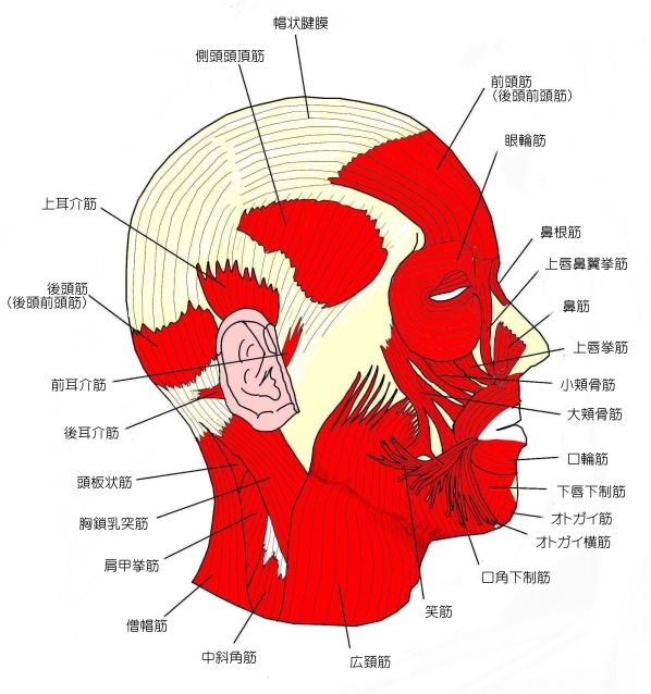
この前頭筋と後頭部に位置する後頭筋を合わせて1つの「 後頭前頭筋 」とし、前頭筋 を 前頭筋腹 ( frontal belly )、そして 後頭筋 を 後頭筋腹 ( occipital belly ) としている資料も見られる。いずれにしても両者は 帽状腱膜 ( epicranial aponeurosis、扁平な腱 ) に移行している。
![]()
【 対性?無対性? 】
「 日本人体解剖学 」の解説文には「 無対性 」という解説になっているが、「 Rauber-Kopsch解剖学 」の解説文では「 左右の前頭筋 」という文言が見られる。また、インターネットで画像検索をかけると、前頭筋を左右分けて描いているイラストを掲載しているサイトは多い。中には、「 上部はしっかりと左右分かれているが、下部においては1つになっている 」というイラストも見られる。
以下は「Wikipedia」の解説の「 structure 」の項の一部を箇条書きに要約したものとなる。
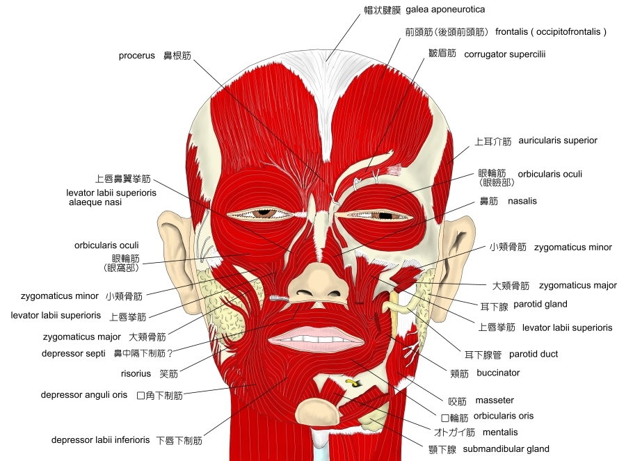
・ 前頭筋は薄く、四辺形で浅筋膜と密着している。
・ 喉頭筋よりも幅広で、その筋線維は寄りより長く薄い色をしている。
・ 骨には付着していない。
・ 内側部の筋線維は 鼻根筋 (procerus) の延長となる。
・ 中間部の筋線維は 皺眉筋 (corrugator muscle) および眼輪筋と交わり まぶた (eyebrow) の皮膚にまで達する。
・ 外側部の筋線維は前頭骨の頬骨突起上で眼輪筋と交わる。
・ 冠状縫合 (colonal suture) の手前で帽状腱膜に移行する。
・ 鼻根部にて左右の前頭筋は癒合している。
以下は「 Rauber-Kopsch解剖学 」の解説文となる。
「 前頭筋は眉や眉間のあたりの皮膚や結合組織から起る.そのさいこの筋は眼瞼の輪走筋を所々で貫き,この輪走筋に対して前頭筋は1つの放射状筋となっていて,さらに眉間下制筋,籔眉筋ならびに眉頭下制筋の筋束をも貫くのである.その線維は頭頂部に向って散開してゆき,両側の前頭結節の高さで弓状にまがり帽状腱膜に移行する.左右の前頭筋の内側縁は,下の方では多少の差はあるがたがいに合しているが,上の方では離れて,そのあいだでは前頭面の一部がこの筋に被われていない.
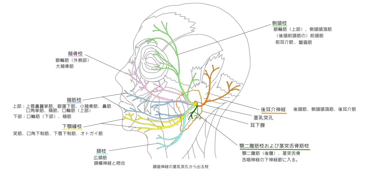
神経支配 : 顔面神経の側頭前頭枝による.
作 用 : この筋は前頭の皮膚に横の皺を寄せ,眉を引きあげる.
変 異 : この筋が欠けていることがある.両側の篩が正中線で交叉しそいることがある.しばしばこの筋がいくつかの筋束に分れている.後頭筋と直接に続くことは極めてまれである. 」

|
 |
|
![]()
【 起 始 】: 鼻根、内眼角、眉間の皮膚
【 停 止 】: 帽状腱膜
「 眉を挙げ額の皮膚に横の皺をつくり、あるいは帽状腱膜と頭皮とを前方に引く。」( 日本人体解剖学 )
・ 神 経 : 顔面神経 の 側頭枝
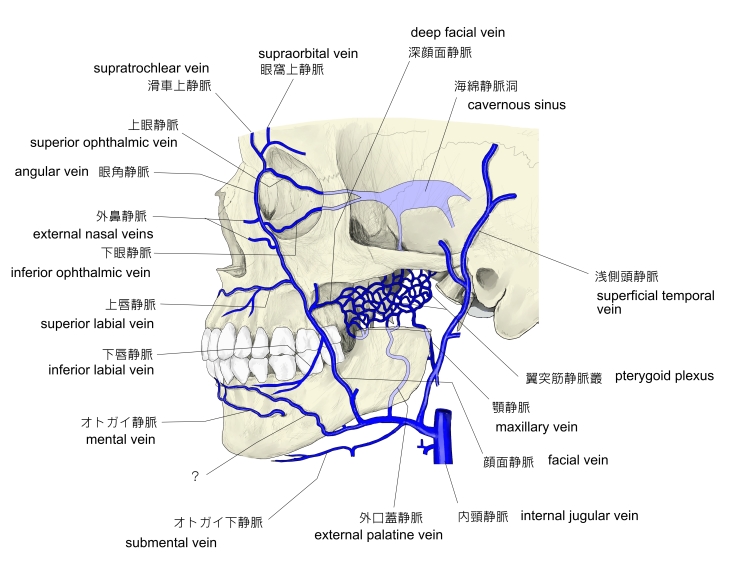
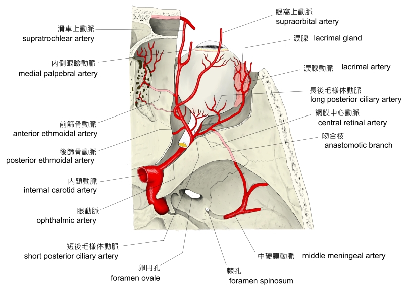
・ 動 脈 :「 receives blood from the supraorbital and supratrochlear arteries 」 ( Wikipedia )
・ 静 脈 : 上記の動脈の説明から静脈も同名のもの( 眼窩上静脈、滑車上静脈 )と思われるがそれに言及している資料
は見当たらない。
|
 |
 |
The frontalis muscle ( from Latin, meaning 'frontal muscle' ) is a muscle which covers parts of the forehead of the skull. Some sources consider the frontalis muscle to be a distinct muscle. However, Terminologia Anatomica currently classifies it as part of the occipitofrontalis muscle along with the occipitalis muscle.[2]
In humans, the frontalis muscle only serves for facial expressions.[3]
The frontalis muscle is supplied by the facial nerve[4] and receives blood from the supraorbital and supratrochlear arteries.
The frontalis muscle is thin, of a quadrilateral form, and intimately adherent to the superficial fascia. It is broader than the occipitalis and its fibers are longer and paler in color. It is located on the front of the head.
The muscle has no bony attachments. Its medial fibers are continuous with those of the procerus ; its intermediate fibers blend with the corrugator and orbicularis oculi muscles, thus attached to the skin of the eyebrows ; and its lateral fibers are also blended with the latter muscle over the zygomatic process of the frontal bone.
From these attachments the fibers are directed upward, and join the galea aponeurotica below the coronal suture.
The medial margins of the frontalis muscles are joined together for some distance above the root of the nose ; but between the occipitales there is a considerable, though variable, interval, occupied by the galea aponeurotica.
In humans, the frontalis muscle only serves for facial expressions.[3]
In the eyebrows, its primary function is to lift them (thus opposing the orbital portion of the orbicularis), especially when looking up. It also acts when a view is too distant or dim.[5]
【 語 句 】
・ distinct : 別個の、明瞭な ・ occipitofrontalis muscle : 後頭前頭筋 ・ occipitalis muscle : 後頭筋 ・ supraorbital artery : 眼窩上動脈 ・ supratrochlear artery : 滑車上動脈 ・ quadrilateral : 四辺形の ・ intimately : 親密に ・superficial fascia : 浅筋膜 ・ pale : 薄い色の ・ procerus : 鼻根筋 ・ corrugator muscle : 皺眉筋 ・ orbicularis oculi muscle : 眼輪筋 ・ zygomatic process of the frontal bone : 前頭骨の頬骨突起 ・ galea aponeurotica: 帽状腱膜 ・ coronal suture : 冠状縫合 ・ eyebrows : まゆ ・ dim : ぼんやりした
![]()