

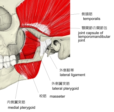
側頭筋 ( そくとうきん、 英 : temporalis muscle )
・ 概 要 |
・ 作 用 |
・ イラスト掲載サイ |
|
・ イラスト |
・ 神経 / 脈管 |
||
・ 起始 / 停止 |
・ Wikipedia |
![]()
・ 「 側頭筋よりも浅層には側頭頭頂筋が存在する。」 ( 日本人体解剖学 )
・ 「 付着腱は上方へ伸びて筋肉内にまで達する。 」 (船戸和弥のHP)
・ 「 純粋な“ 咬むための筋 ”である。 」 (船戸和弥のHP)
以下は「 船戸和弥のHP 」の解説文となる。
「 側頭筋は扇状になって側頭窩および側頭筋膜から起始する。筋線維は収斂して、頑丈な腱をもって下顎骨筋突起に付着する。付着腱は上方へ伸びて筋肉内にまで達する。側頭筋は頬骨弓下を通過して、その付着部に達する。その筋線維がかなりの長さであるので、筋はかなりの収縮可能性を有するし、かつ純粋な“咬むための筋”である。歯をかみ合わせると、側頭筋の収縮を耳介の上方で触れることができる。側頭部をコメカミというのは、コメをカムときに動くからである。」
 |
 |
|
 |
 |
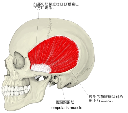
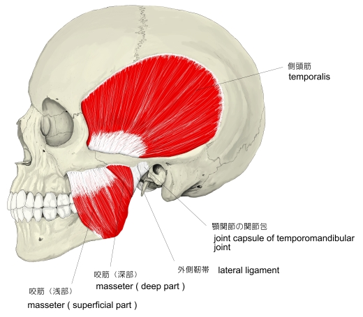
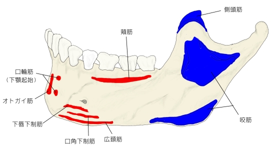
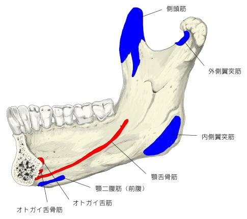
【 起 始 】: 側頭骨 の側頭全面と 側頭筋膜 の内側面から起始し、下前方に向かって筋線維が集まるように走る。
【 停 止 】: 下顎骨 の 筋突起とそれに続く 下顎枝 の内側面 「 日本人体解剖学 」
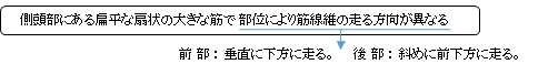
下顎を引き上げ( 前部の筋線維 )、なおかつ後方に引く。( 後部の筋線維 )
 |
|
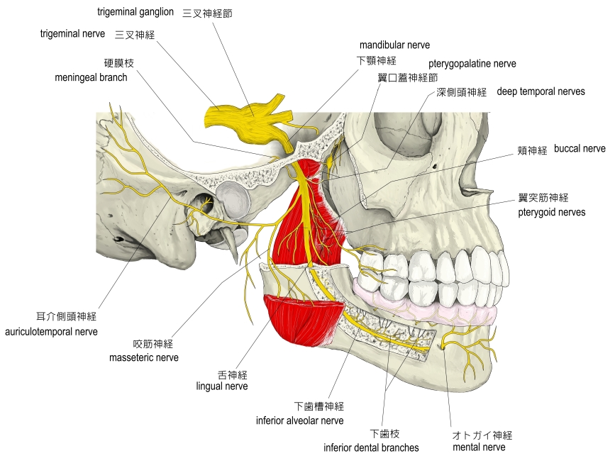
・ 神 経 : 下顎神経( 三叉神経 の第3枝 )の枝の 深側頭神経
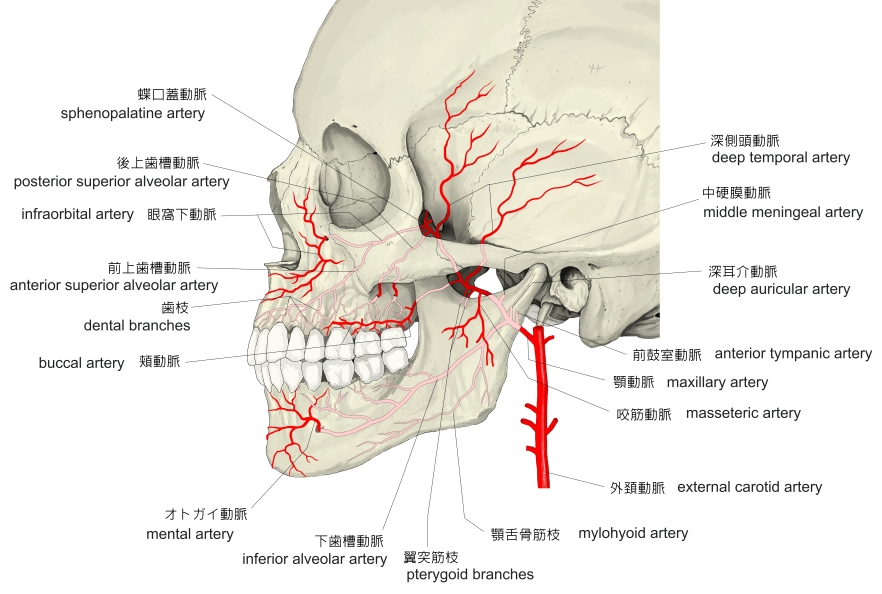
・ 動 脈 : 深側頭動脈 「 Wikipedia 」
・ 静 脈 : 上記の動脈より同名の 深側頭静脈 と思われるが、それに言及している資料は見当たらない。
|
|
「 歯をかみ合わせると、側頭筋の収縮を耳介の上方で触れることができる。側頭部をコメカミというのは、コメをカムときに動くからである。」(船戸和弥のHP)
The temporal muscle, also known as the temporalis, is one of the muscles of mastication. It is a broad, fan-shaped muscle on each side of the head that fills the temporal fossa, superior to the zygomatic arch so it covers much of the temporal bone.[1]
In humans, it arises from the temporal fossa and the deep part of temporal fascia. It passes medial to the zygomatic arch and forms a tendon which inserts onto the coronoid process of the mandible, with its insertion extending into the retromolar fossa posterior to the most distal mandibular molar.[2] In other mammals, the muscle usually spans the dorsal part of the skull all the way up to the medial line. There, it may be attached to a sagittal crest, as can be seen in early hominins like Paranthropus aethiopicus.
The temporal muscle is covered by the temporal fascia, also known as the temporal aponeurosis. This fascia is commonly used in tympanoplasty, or surgical reconstruction of the eardrum.
The muscle is accessible on the temples, and can be seen and felt contracting while the jaw is clenching and unclenching.
【 語 句 】
・ mastication : 咀嚼 ・ temporal fossa : 側頭窩 ・ zygomatic arch : 頬骨弓 ・ temporal bone : 側頭骨 ・ temporal fascia : 側頭筋膜 ・ coronoid process of the mandible : 下顎骨の筋突起 ・ retromolar fossa : 臼歯後窩 ・ mandibular molar : 下顎臼歯 ・ sagittal crest : 矢状隆起 ・ hominin : ヒト科? ・ Paranthropus aethiopicus : パラントロプス・エチオピクス ・ temporal aponeurosis : 側頭腱膜 ・ tympanoplasty : 鼓室形成術 ・ eardrum : 鼓膜 ・ temple : こめかみ ・ contract : 引き締める ・ clench : くいしばる
【 Development 】
The temporalis is derived from the first pharyngeal arch in development.
【 Innervation 】
As with the other muscles of mastication, control of the temporal muscle comes from the third ( mandibular ) branch of the trigeminal nerve. Specifically, the muscle is innervated by the deep temporal nerves.
【 Blood supply 】
The muscle receives its blood supply from the deep temporal arteries which anastomose with the middle temporal artery.
The temporal muscle is the most powerful muscle of the temporomandibular joint. The temporal muscle can be divided into two functional parts ; anterior and posterior. The anterior portion runs vertically and its contraction results in elevation of the mandible (closing the mouth). The posterior portion has fibers which run horizontally and contraction of this portion results in retrusion of the mandible.[3]
When lower dentures are fitted, they should not extend into the retromolar fossa to prevent trauma of the mucosa due to the contraction of the temporalis muscle.[2]
【 語 句 】
・ be derived from ~ : ~ に由来する ・ pharyngeal arch : 鰓弓 ・ trigeminal nerve : 三叉神経 ・ deep temporal nerve : 深側頭神経 ・ anastomose with ~ : ~ と吻合する ・ middle temporal artery : 中側頭動脈 ・ temporomandibular joint : 顎関節 ・ retrusion : 下顎の後方移動 ・ denture : 総義歯 ・ trauma : 精神的外傷 ・ mucosa : 粘膜 ・ contraction : 収縮
![]()