・「長さ:約4mm、幅:約2mm」(Wikipedia)

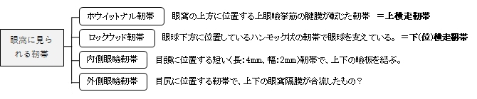
以下は眼窩の前部にある靭帯を記した簡単な表となる。
「日本人体解剖学 (下巻) 」には以下のような解説が見られる。
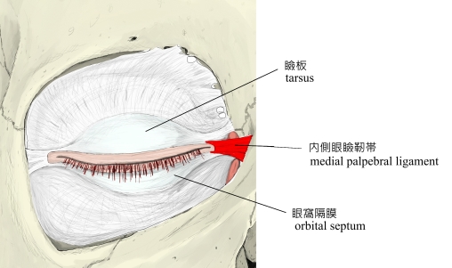
「上・下瞼板は、内側眼瞼靭帯によって結合される。この靭帯は、上顎骨の前頭突起から起こり弓をなして涙嚢の前側にいき、ついでこれを包む前後の2脚に分かれ、前脚は前涙嚢稜に至り、後脚は後涙嚢稜に至る。」
また、「Rauber-Kopsch解剖学」では以下のような解説が見られる。
「内眼角のところでは内側眼瞼靱帯Ligamentum palpebrale nasale(図662, 663)が眼瞼板に結合してその線維構成に移行している.内側眼瞼靱帯は斜めに立っている板で,内眼角から起こって上顎骨の前頭突起にまで伸びて,皮膚の直下で涙嚢の盲端部の前面にある.眼を閉じているときにはその前稜を容易に触知することができる.」
【Wikipedia 】
The medial palpebral ligament (medial canthal tendon) is about 4 mm in length and 2 mm in breadth. Its anterior attachment is to the frontal process of the maxilla in front of the lacrimal groove, and its posterior attachment is the lacrimal bone. Laterally, it is attached to the tarsus of the upper and lower eyelids.
Crossing the lacrimal sac, it divides into two parts, upper and lower, each attached to the medial end of the corresponding tarsus.
As the ligament crosses the lacrimal sac, a strong aponeurotic lamina is given off from its posterior surface; this expands over the sac, and is attached to the posterior lacrimal crest.
語句
・frontal process:前頭突起 ・maxilla:上顎骨 ・lacrimal groove:涙嚢溝 ・lacrimal bone:涙骨 ・tarsus:瞼板 ・eyelid:まぶた ・aponeurotic:腱膜の ・lamina:薄膜/板 ・posterior lacrimal crest:後涙嚢稜
【 他の参考となるサイト 】
・イラストを掲載しているサイトⅠ
・イラストを掲載しているサイトⅡ
・イラストを掲載しているサイトⅢ
・イラストを掲載しているサイトⅣ
・イラストを掲載しているサイトⅤ