関節上腕靭帯とは

「船戸和弥のホームページ」では以下のように解説している。
「関節上腕靱帯は関節包が周囲の筋の腱で補強されている以外の所で、主に深層の線維の集団で肥厚する部である。特に前壁を内面から見るときに他の部と判別できる。関節唇の上、前、および下部から出て不明瞭な線維束となり、解剖頚に着く。上にあるもの(上束)と前壁を斜めに外下方に走るもの(前束)との間の前壁外上部は関節包の弱いところとなる。ここが肩甲下筋腱下包と交通しているとき、その孔はWeitbrechtの卵円孔と呼ばれることがある。上腕を外転すると下束と前束が緊張し、回外すると上束、前束、下束が緊張する。」
また、以下は「Wikipedia」の解説文となる。
「In human anatomy, the glenohumeral ligaments (GHL) are three ligaments on the anterior side of the glenohumeral joint (i.e. between the glenoid cavity of the scapula and the head of the humerus; colloquially called the shoulder joint). Reinforcing the anterior glenohumeral joint capsule, the superior, middle, and inferior glenohumeral ligaments play different roles in the stability of the head of the humerus depending on arm position and degree of rotation.[1]
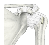
Location:The ligaments may be best seen by opening the capsule at the back of the joint and removing the head of the humerus:[2]
- One on the medial side of the joint passes from the medial edge of the glenoid cavity to the lower part of the lesser tubercle of the humerus.
- A second at the lower part of the joint extends from the under edge of the glenoid cavity to the under part of the anatomical neck of the humerus.
- A third at the upper part of the joint is fixed above to the apex of the glenoid cavity close to the root of the coracoid process, and passing downward along the medial edge of the tendon of the Biceps brachii, is attached below to a small depression above the lesser tubercle of the humerus.
In addition to these, the capsule is strengthened in front by two bands derived from the tendons of the Pectoralis major and Teres major respectively.[2]
Function:During abduction of the arm the middle and inferior ligaments become taut while the superior ligament relaxes. The radius of curvature of the head of the humerus is greater superiorly than inferiorly, which further stretches these ligaments so that they keep the articular surfaces of the joint in their close-packed position. [3]
During abduction the greater tubercle of the humerus comes in contact with the upper margin of the glenoid cavity, which limits maximum abduction. By rotating the humerus laterally, this contact is delayed because the greater tubercle is pulled back so that the bicipital groove faces the coracoacromial ligament. This slightly slackens the inferior fibres of the glenohumeral ligament, allowing an abduction of 90°. Combining abduction with 30° flexion in the plane of the scapula causes a delay in the tightening of the ligament resulting in a maximum abduction of 110°. [3]
During rotation of the arm lateral rotation stretches all three ligaments while medial rotation relaxes them. [3]」
【 他の参考となるサイト 】
・イラストや写真を掲載しているサイト-Ⅰ
・イラストや写真を掲載しているサイト-Ⅱ
・イラストや写真を掲載しているサイト-Ⅲ
・イラストや写真を掲載しているサイト-Ⅳ(肩関節の関節窩を外側から見たもの)


