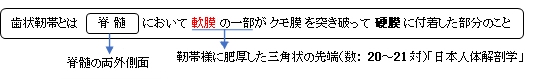
歯状靭帯 ( しじょうじんたい、英:denticulate ligament )
働き:髄膜内における脊髄位置の保持 ・「前根と後根の間に張って軟膜と硬膜とを連絡する。」(日本人体解剖学) 以下は「Wikipedia」の解説文の一部を箇条書きとしたものとなる。 1. 頚髄と胸髄に見られる。 2. それぞれの靭帯が18から20の三角状の線維束(?)より成る。 3. 頸部、胸部でそれぞれ異なる特徴を有する。 ・ 頸部 : 胸部より短く(3~5mm)、数が多い。 ・ 胸部 : 下部になると長さが21から26mmとなる。
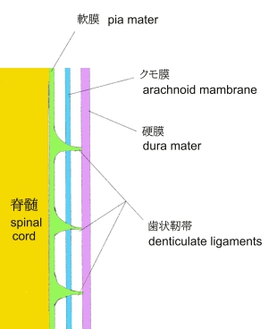
以下は「日本人体解剖学」の「脊髄軟膜」の解説文となる。 「薄い結合組織性の膜で、脊髄の表面を直接多い、血管を伴ってその溝・裂よび実質中に侵入する。脊髄の前正中裂に入るものを前縦中隔という。なお、軟膜は脊髄の側面では靭帯様に肥厚し、これから出る三角形の小突起は、前根と後根の間に張って軟膜と硬膜とを連絡する。その数は20~21対あって脊髄を支持し、形が鋸歯状で歯状靭帯と呼ばれる。 」
The denticulate ligaments, also known as dentate ligaments, are triangular shaped ligaments that anchor the spinal cord along its length, at each side, to the dura mater. The bases of the ligaments arise in the pia mater and they are firmly attached to the arachnoid mater and dura mater at the apex. They have 21 attachments per side. Their tooth-like appearance originates the word which derives from Latin denticulatus, from denticulus ‘small tooth’. The denticulate ligaments are traditionally believed to provide stability for the spinal cord against motion within the vertebral column. 【Structure】 Each denticulate ligament is composed of a single narrow fibrous strip that extends from the craniovertebral junction to T12. Each ligament features 18-20 triangular extensions that attach to the dura at their apices. The triangular extensions are smaller and more numerous at the cervical levels, and are larger and less numerous at the thoracic levels. The apices of the extensions attach to the dura via fibrous bands at cervical levels (each band 3–5 mm (0.12–0.20 in) long) and lower thoracic levels (21–26 mm (0.83–1.02 in) long), whereas they attach directly to the dura at upper thoracic levels. 【Mechanical properties】 Denticulate ligaments are characterised by high extensibility (on average 50% of their initial length) and relatively low force necessary to rupture them (around 1 N). Their strength, especially in cervical region, decreases in caudal orientation. 【 語 句 】 ・spinal cord:脊髄 ・dura mater:硬膜 ・pia mater:軟膜 ・arachinoid mater:クモ膜 ・vertebral column:脊柱 ・lumbar spinal stenosis:腰部脊柱管狭窄症 ・disc herniations:椎間板ヘルニア ・facet hypertrophy:面?肥大 ・ligamentum flavum hypertrophy:黄色靭帯肥大? ・degenerative joint disease:変性関節疾患 ・osteophyte:骨棘、骨増殖体 ・craniovertebral:頭蓋脊椎の ・feature:特色をなす ・cervica頸部の ・thoracic:胸部の ・apice:先端 ・abundant:豊富な ・affect:影響を及ぼす、作用する ・alter:変更する
|