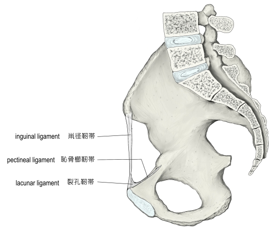
鼡径靭帯とは
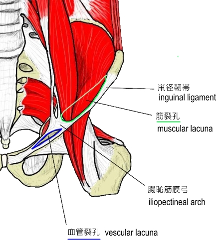
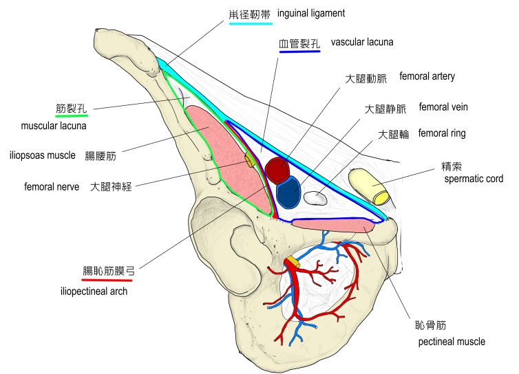
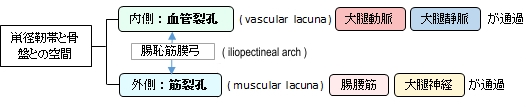
また、鼡径靭帯と骨盤の間の空間は、腸恥筋膜弓という帯状の線維組織で内・外の2つに分けられる。その内側のものを血管裂孔、そして外側のものを筋裂孔といい、血管や筋肉、そして神経などが通過するところとなっている。
以下は「ウィキまとめ」の解説文となる。
「《同義語》プーパル靱帯Poupart's ligament
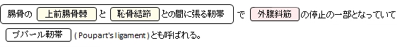
上前腸骨棘*と恥骨結節との間に張る靱帯で外腹斜筋の停止腱の作る腱弓が発達したもの.皮膚上から容易に触れることができる.この鼡径靱帯と恥骨上枝との間の間隙は腸恥筋膜弓によって筋裂孔と血管裂孔に2分される.筋裂孔は腸腰筋と大腿神経が通り,血管裂孔は大腿動静脈とリンパ管が通る.鼡径靱帯の内側部は後方に向かって広がり恥骨筋膜と合し裂孔靱帯となって恥骨櫛内側部に至る.これは血管裂孔の外側縁をなし,この線維のつづきが恥骨櫛に沿って延びているものを恥骨櫛靱帯という.
裂孔靱帯のつづきとして腹直筋鞘を作る内腹斜筋腱膜の前面に向かって出る線維が反転靱帯reflex ligamentであり浅鼡径輪の後方に位置し,鼡径管*の下壁の一部を作る(Francois Poupartはフランスの解剖学者,1661-1708). 」
また、以下は「Wikipedia」の解説文となる。
「The inguinal ligament (Poupart's ligament) is a band running from the pubic tubercle to the anterior superior iliac spine. Its anatomy is very important for operating on hernia patients.
Structure:It forms the base of the inguinal canal through which an indirect inguinal hernia may develop.
The inguinal (crural) ligament runs from the anterior superior iliac spine of the ilium to the pubic tubercle of the pubic bone. It is formed by the external abdominal oblique aponeurosis and is continuous with the fascia lata of the thigh.
There is some dispute over the attachments.[1]
Structures that pass deep to the inguinal ligament include:
The midpoint of inguinal ligament is midpoint between the anterior superior iliac spine and pubic tubercle.[citation needed]
Function: The ligament serves to contain soft tissues as they course anteriorly from the trunk to the lower extremity. This structure demarcates the superior border of the femoral triangle.[2] It demarcates the inferior border of the inguinal triangle.
The midpoint of inguinal ligament is halfway between the anterior superior iliac spine and pubic tubercle. It's the landmark for the femoral nerve. The mid inguinal point is halfway between the anterior superior iliac spine and pubic symphysis. It's the landmark for femoral artery.」
【イラスト】
・参考となるイラストを掲載しているサイト-Ⅰ
・参考となるイラストを掲載しているサイト-Ⅱ
・参考となるイラストを掲載しているサイト-Ⅲ