後縦靭帯とは

以下は、「船戸和弥のホームページ」の「解剖学」からの引用文となる。
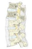
「 後縦靱帯は椎体と椎間円板の後面に沿い、脊柱管の前壁を縦走する。上端は幅が広く、大後頭孔前縁より約1cm上方の斜台から起こり(この部の深層の部は蓋膜と呼ばれる)、下方ほど狭くなって仙骨管の前壁に達する。椎間円板およびそれに接する椎体の縁と固く結合し、椎体後面の中部とは結合がゆるい。とくにひゅそうの線維は4~5個の椎体を越えて椎間円板から椎間円板へと結合し、この靱帯が椎間円板に着くところでは特に幅が広くなっている。前・後縦靱帯は椎体と椎間円板からなる柱を前後から支えるとともに、椎間円板の弾性によって緊張させられている。 」
また、以下は「Wikipedia」の解説文となる。
「The posterior longitudinal ligament is situated within the vertebral canal, and extends along the posterior surfaces of the bodies of the vertebræ, from the body of the axis, where it is continuous with the membrana tectoria, to the sacrum.[1]
It is broader above than below, and thicker in the thoracic than in the cervical and lumbar regions. The ligament is more narrow at the vertebral bodies and wider at the intervertebral disc space which is more pronounced than the anterior longitudinal ligament. This is significant in understanding certain pathological conditions of the spine such as the typical location for a spinal disc herniation.
In the situation of the intervertebral fibrocartilages and contiguous margins of the vertebræ, where the ligament is more intimately adherent, it is broad, and in the thoracic and lumbar regions presents a series of dentations with intervening concave margins; but it is narrow and thick over the centers of the bodies, from which it is separated by the basivertebral veins.
This ligament is composed of smooth, shining, longitudinal fibers, denser and more compact than those of the anterior ligament, and consists of superficial layers occupying the interval between three or four vertebræ, and deeper layers which extend between adjacent vertebræ.
It functions to prevent hyperflexion of the vertebral column.」
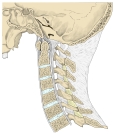
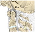
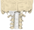
【 イラスト 】
【 他の参考となるサイト 】
・後縦靭帯のイラストや写真を掲載しているサイト①
・後縦靭帯のイラストや写真を掲載しているサイト②
・後縦靭帯のイラストや写真を掲載しているサイト③
・後縦靭帯のイラストや写真を掲載しているサイト④
・ほぼ全体が分かるイラストを掲載しているサイト⑤